
Obesity-Related Mitochondrial Mitophagy & Damage Detection Service
InquiryMetabolic disorders like obesity put heavy strain on cellular energy networks, especially in key metabolic tissues such as white and brown adipose tissue, skeletal muscle, and the liver. Overfeeding and high-fat exposure lead to nutrient oversupply, which increases reactive oxygen species (ROS) production, disrupts mitochondrial membrane potential, and triggers structural fragmentation. Under normal conditions, cells clean up damaged organelles using mitophagy—a selective autophagic pathway that sequesters dysfunctional mitochondria into autophagosomes for lysosomal degradation.
Preclinical Mitophagy Assays: Quantifying Obesity-Induced Mitochondrial Damage
Understanding these complex mitochondrial pathways requires sensitive, reliable quantitative tools tailored for preclinical research. Protheragen provides comprehensive, high-throughput preclinical assay platforms specifically designed to analyze obesity-induced mitochondrial damage and track mitophagy dynamics. Our goal is to help drug discovery teams validate therapeutic targets, evaluate small molecules, and map metabolic interventions without the friction of unstandardized testing.
Core Technologies
Accurately evaluating mitophagy and organelle health requires multi-parametric assay systems that capture dynamic morphological and biochemical changes across primary cultures and in vivo tissue models.
We utilize advanced biosensor models including mt-Keima and Rosella constructs, alongside Cox8-GFP-mCherry tandem reporters. The mt-Keima assay uses a pH-sensitive fluorescent protein targeted to the mitochondrial matrix. In acidic lysosomal environments after autophagic engulfment, its excitation spectrum shifts dramatically, allowing precise quantification of mitophagy flux via flow cytometry or confocal imaging.
Mitophagy occurs through distinct ubiquitin-dependent and ubiquitin-independent pathways. We run specialized biochemical assays targeting the PINK1-Parkin cascade, measuring PINK1 stabilization on the outer membrane, Parkin recruitment, and ubiquitin phosphorylation at Ser65. For ubiquitin-independent routes, we quantify membrane receptors such as BNIP3, NIX (BNIP3L), and FUNDC1 through automated Western blot, capillary immunoassay, and high-content screening platforms.
Assessing organelle integrity requires evaluating structural breakdown alongside autophagic removal. Our panel tracks key damage markers including mitochondrial permeability transition pore (mPTP) opening, cyto-nuclear DNA ratio (mtDNA copy number), and outer membrane protein loss (e.g., TOMM20, VDAC1 degradation) relative to inner matrix markers like CS and COX IV.
We integrate metabolic bioenergetics with mitophagy assays. Using high-resolution respirometry and extracellular flux analysis, our platform measures oxygen consumption rate (OCR), ATP production, proton leak, and maximal respiratory capacity alongside live-cell superoxide and hydrogen peroxide tracking.
Service Scope
Protheragen offers a flexible, full-service preclinical testing framework designed to evaluate mitochondrial health in metabolic Disease Models.
- In Vitro Mitophagy Flux Quantification
Real-time tracking of autophagic flux in primary adipocytes, myotubes, and hepatocytes treated with fatty acids (e.g., palmitate), high glucose, or inflammatory cytokines.
- Ex Vivo & Tissue-Specific Mitophagy Assays
Microscopic and biochemical evaluation of organelle clearance in metabolic tissues isolated from diet-induced obesity (DIO) or genetically modified rodent models.
- Mitochondrial Damage & Membrane Potential Profiling
High-throughput screening using JC-1, TMRM, and fluorescent indicators to measure membrane potential loss and organelle depolarization.
- Organelle Dynamics & Morphology Analysis
Automated high-content imaging to measure mitochondrial fission and fusion dynamics (Drp1, Mfn1/2, Opa1 tracking).
Screening small molecules, natural products, or biologic candidates for their ability to restore impaired mitophagy or mitigate excessive organelle degradation.
Custom multiplex gene and protein expression panels evaluating upstream autophagic regulators (ULK1, Beclin1, LC3-II/I ratios, p62/SQSTM1 clearance).
Accelerate your metabolic research by partnering with Protheragen
Workflow
Our streamlined preclinical service process delivers reliable, high-quality data while keeping your project timeline on track.
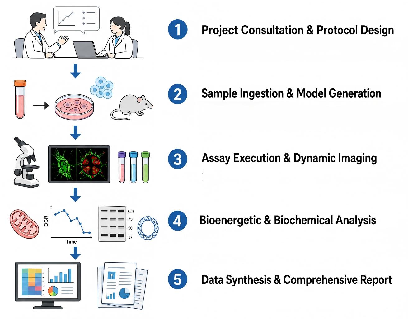
- Step 1: Define study objectives, select optimal cell or tissue models, and customize experimental protocols.
- Step 2: Process client samples or induce metabolic stress in target cell lines and animal models.
- Step 3: Apply ratiometric biosensors, high-content imaging, and fluorometric probes to track live-cell mitophagy.
- Step 4: Quantify oxygen consumption rates, protein phosphorylation, mtDNA integrity, and organelle clearance markers.
- Step 5: Normalize experimental readouts, run statistical analysis, and deliver clear, publication-ready research reports.
Fields of Application
Our specialized preclinical mitophagy and mitochondrial damage detection services directly support research across several key therapeutic areas and metabolic disease models.
- Anti-Obesity Drug Discovery: Validating therapeutic compounds aimed at reducing fat accumulation by enhancing mitochondrial quality and oxidative capacity in brown and beige fat.
- Type 2 Diabetes & Insulin Resistance: Evaluating candidates that reduce glucolipotoxicity-induced mitochondrial damage in skeletal muscle and liver cells.
- Non-Alcoholic Fatty Liver Disease (NAFLD/MASH): Assessing compounds designed to clear damaged hepatic mitochondria, lowering intracellular oxidative stress and liver inflammation.
- Metabolic Cardiomyopathy: Evaluating protective therapies that stop excessive or defective mitophagy in heart tissue exposed to high lipid environments.
- Nutraceutical & Lifestyle Research: Testing dietary compounds, functional foods, and exercise-mimetic formulations for their impact on mitochondrial quality control.
Advantages
Partnering with Protheragen provides your drug discovery team with specialized preclinical expertise, validated multi-parametric assay platforms, and rapid turnaround times tailored to metabolic research.
Tailored Preclinical Focus
At Protheragen, we concentrate purely on preclinical testing. This specialized focus allows us to optimize cell models, ex vivo assays, and tissue profiling without the high overhead or complex protocols of clinical service providers.
Validated Multi-Reporter Platforms
Our assays use dual-confirmation systems—combining live-cell ratiometric imaging with capillary westerns—to eliminate false positives caused by general autophagy or lysosomal dysfunction.
High-Throughput Assay Optimization
Automated screening pipelines let us test broad compound libraries quickly, helping drug discovery teams save weeks during early hit-to-lead phases.
Context-Specific Primary Models
We maintain optimized primary cell cultures (including rodent and human adipocytes, hepatocytes, and skeletal myotubes) exposed to physiologically relevant metabolic stress, delivering data that reflects true tissue biology better than immortalized lines.
Contact Our Team for More Information and to Discuss Your Project
Publication Data
Title: LncRNA H19 governs mitophagy and restores mitochondrial respiration in the heart through Pink1/Parkin signaling during obesity.
Journal: Cell Death Dis, 2021
DOI: https://doi.org/10.1038/s41419-021-03821-6
Summary: This study reveals lncRNA H19 curbs excessive Pink1/Parkin-mediated mitophagy to restore cardiac mitochondrial respiration in obesity. Palmitic acid boosts Dnmt3b-driven H19 promoter methylation, repressing H19. H19 sequesters eIF4A2 to block Pink1 mRNA translation, relieving obese heart injury.
Key Findings
- Obesity-related cardiac phenotype: In palmitic acid-treated H9c2 cardiomyocytes and Lep-/- obese mice, lncRNA H19 expression drops, alongside impaired mitochondrial respiration, reduced mitochondrial mass and hyper-activated mitophagy.
- H19 protective function: Cardiomyocyte-specific H19 overexpression rescues mitochondrial respiratory capacity, recovers mitochondrial quantity and improves diastolic cardiac function under obese stress.
- Mitophagy pathway identification: Excessive mitophagy is predominantly driven by the Pink1/Parkin axis, not Fundc1 or Bnip3/Nix; Pink1 knockdown reverses palmitic-acid-provoked respiratory defects.
- Post-translational regulatory mechanism: H19 binds translation factor eIF4A2, preventing eIF4A2 association with Pink1 mRNA and suppressing Pink1 protein synthesis without altering Pink1 mRNA abundance.
- Epigenetic upstream control: Palmitic acid elevates Dnmt3b expression and activity, increasing CpG methylation on the H19 promoter and silencing H19 transcription.
- Pathophysiological note: Excessive, rather than insufficient, mitophagy mediates cardiomyocyte damage in early-stage obesity.
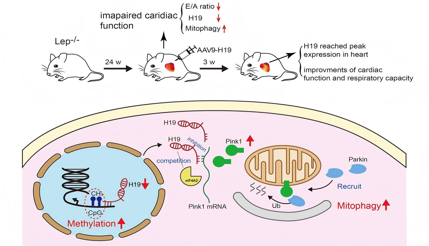 Fig.1 Mechanism diagram: How lncRNA H19 controls mitophagy to protect the obese heart. (Wang, et al.; 2021)
Customer Review
Accelerating Small Molecule Lead Optimization
"Protheragen provided exceptional support during our lead optimization campaign for a novel mitochondrial-targeted therapeutic. Their mt-Keima flow cytometry and bioenergetic assays gave us clear, reproducible data on mitophagy restoration in palmitate-stressed hepatocytes. Their technical team helped refine our assay parameters, and the quick turnaround kept our drug discovery timeline moving forward without delay."
Dr. A. T., Director of Pharmacology
Standardizing Ex Vivo Tissue Profiling
"Their Seahorse respirometry and mitochondrial isolation protocols were spot on for our project. We had been running into consistency issues trying to measure ROS in isolated visceral adipocytes, but Protheragen stepped in and delivered reliable, reproducible data right on schedule. The technical team was super accessible and easy to collaborate with throughout the study."
Dr. E. R., Principal Investigator
Frequently Asked Questions
-
How do your assays distinguish between selective mitophagy and general macroautophagy?
We use organelle-targeted fluorescent probes (like mt-Keima and Cox8-GFP-mCherry) that specifically report when mitochondria enter acidic lysosomes. Combining these readouts with specific outer mitochondrial membrane protein degradation assays ensures we measure true selective organelle clearance rather than non-specific bulk autophagy.
-
Can Protheragen evaluate mitophagy in primary human tissue samples?
Yes, we process primary human preadipocytes, hepatocytes, and muscle progenitors, culturing and differentiating them under metabolic stress conditions. We also analyze frozen or freshly isolated tissue biopsies provided by your team using our validated ex vivo assay protocols.
-
What makes your high-fat diet (HFD) tissue profiling different from standard assays?
We use multi-parametric tissue processing protocols that isolate intact, functional mitochondria alongside whole-tissue lysates. This approach lets us measure bioenergetic function, structural damage, and autophagosome engulfment in parallel from the same tissue batch.
-
Do you offer drug library screening services using these assays?
Absolutely. Our high-content imaging platforms are scaled for 96-well and 384-well formats, making them ideal for screening small molecule libraries, biologics, or natural product derivatives to identify mitophagy modulators.
-
Why is measuring mitochondrial membrane potential (ΔΨm) alone insufficient to assess mitophagy?
Membrane depolarization is an early step in mitochondrial damage, but it doesn't guarantee that autophagic degradation will follow. In some diseased tissues, depolarized organelles accumulate without being cleared. Our assays track the entire pathway—from initial depolarization to final lysosomal destruction.
-
What controls do you include to confirm lysosomal turnover of mitochondria?
We regularly use lysosomal inhibitors (such as bafilomycin A1 or chloroquine) alongside tested compounds. Blocking lysosomal acidification helps us verify whether changes in signal reflect increased autophagic flux or blocked downstream degradation.
-
Can these services support regulatory filings for preclinical candidates?
Yes. All assays follow strict standard operating procedures (SOPs), complete with comprehensive data trails, precise validation metrics, and structured reporting designed to support regulatory submissions and IND packages.
-
What primary cell models work best for obesity-related studies?
Differentiated 3T3-L1 adipocytes, primary rodent or human subcutaneous/visceral adipocytes, C2C12 myotubes, and primary human hepatocytes (PHH) exposed to fatty acid mixtures (like oleate/palmitate) provide highly reliable, biologically relevant models.
-
How do you handle challenging tissue samples with high fat content?
Adipose tissue requires specialized homogenization and lipid-separation protocols. Our lab utilizes optimized centrifugation and buffer systems designed to remove lipid layers cleanly, preventing sample interference during Western blots and fluorometric assays.
Contact Us
Protheragen delivers high-quality, quantitative preclinical assay solutions to advance your metabolic research. Whether you need targeted hit identification, lead optimization, or detailed ex vivo tissue profiling, our team is ready to support your scientific goals. We invite you to Contact Protheragen for more information about our services.
Reference
- Wang, S.H.; et al. LncRNA H19 governs mitophagy and restores mitochondrial respiration in the heart through Pink1/Parkin signaling during obesity. Cell Death Dis. 2021, 12, 557. (CC BY 4.0)
All of our services and products are intended for preclinical research use only and cannot be used to diagnose, treat or manage patients.