
Obesity-Related Mitochondrial Morphology & Structural Analysis Service
InquiryObesity is no longer understood simply as an accumulation of excess adipose tissue. Modern metabolic research shows that it is deeply rooted in severe cellular energy defects. At the heart of this metabolic breakdown is the mitochondrion. Chronic nutrient overload and elevated circulating free fatty acids induce severe lipotoxicity, which directly disrupts the delicate balance of mitochondrial dynamics. Instead of maintaining healthy, elongated networks capable of efficient beta-oxidation, mitochondria in obese tissues frequently shift toward excessive, uncontrolled fission. This structural fragmentation leads to a dramatic drop in ATP synthesis, elevated reactive oxygen species (ROS) production, systemic insulin resistance, and the activation of chronic low-grade inflammatory pathways.
Advanced Mitochondrial Morphology Screening Services for Preclinical Obesity Research
Understanding exactly how obesity shifts mitochondrial architecture is critical for developing effective metabolic therapeutics. Protheragen provides a specialized obesity-related mitochondrial morphology & structural analysis service designed specifically for preclinical drug discovery and mechanistic studies. By coupling advanced high-resolution imaging with automated morphometric quantification, we enable researchers to map alterations in mitochondrial network complexity, volume, and spatial distribution across various metabolic cell and tissue models. Our platform translates highly complex structural phenotypes into actionable, reproducible datasets, helping you confidently validate therapeutic targets and assess drug candidates.
Core Technologies
To accurately capture the structural heterogeneity of compromised mitochondrial networks, our platform integrates several advanced imaging and quantitative technologies:
Enables deep optical sectioning and crisp 3D reconstruction of intricate mitochondrial networks within intact cells and tissues, minimizing out-of-focus fluorescence.
Breaks the optical diffraction limit to reveal sub-diffraction structures, such as individual mitochondrial cristae alignment and subtle outer-membrane remodeling events.
Utilizing advanced machine-learning algorithms, our software automatically extracts critical structural descriptors—including aspect ratio, form factor, circularity, branching index, and total network length—eliminating operator bias.
Designed to track real-time mitochondrial structural transitions, structural velocity, and morphological remodeling in response to metabolic stressors or candidate compounds over extended periods.
Service Scope
Our specialized service scope is split into two complementary assessment pillars designed to give you a complete picture of structural and kinetic changes in Metabolic Disease Models.
This service is optimized to visualize and quantify the spatial layout, overall abundance, and structural state of mitochondrial networks in cells under lipotoxic stress. We use a combination of advanced fluorescent probes (such as Mitotracker dyes, TMRE, and genetically encoded matrix-targeted fluorescent proteins) to tag mitochondria in various preclinical configurations.
Our quantitative pipeline assesses structural traits like fragmentation status, network branching, and absolute mitochondrial volume. This allows you to evaluate how effectively your test compounds rescue fragmented, punctate mitochondria and restore them into healthy, interconnected tubular networks. Furthermore, live-cell tracking capabilities allow us to measure structural velocity and look at how individual mitochondrial footprints move and rearrange within insulin-resistant or lipid-loaded cell lineages over time.
Mitochondrial shape is maintained by a continuous balance between fusion and fission events. Obesity often shifts this balance toward fission, driven by the upregulation or hyperactivation of proteins like dynamin-related protein 1 (Drp1) and fission 1 protein (Fis1), while downregulating fusion elements like mitofusins (Mfn1/2) and optic atrophy 1 (Opa1).
This assay service focuses directly on measuring these structural kinetics. We look at the activation, recruitment, and localized assembly of fission and fusion complexes on the outer mitochondrial membrane. Using techniques like photo-activatable green fluorescent proteins (such as mt-PA-GFP diffusion) and customized content-mixing assays, we directly quantify the rate of functional fusion and fission events. This gives you a clear, quantitative look at how your Small Molecules, biologics, or genetic interventions modulate the core machinery governing mitochondrial dynamics.
Request a Technical Consultation with Our Specialists Today
Workflow
Moving a project from initial concept to finalized quantitative metrics requires a highly reproducible, structured sequence that handles fragile metabolic samples with extreme care.
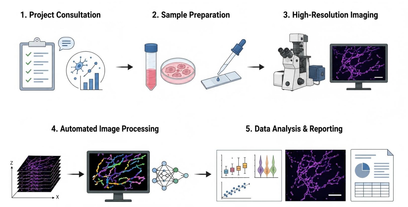
- Step 1: Define specific experimental parameters, custom metabolic models, structural biomarkers, and quantitative endpoints for the study.
- Step 2: Optimize fixation protocols or live-cell fluorescent staining procedures tailored precisely to selected metabolic samples.
- Step 3: Capture detailed, high-dimensional micrographs using advanced confocal or super-resolution platforms under stringent quality controls.
- Step 4: Segment mitochondrial networks using specialized machine-learning algorithms to ensure unbiased structural parameter extraction.
- Step 5: Compile comprehensive structural metrics, statistical evaluations, and representative high-quality imagery into client-ready data packages.
Fields of Application
Our morphological and structural analysis services support multiple areas of preclinical drug discovery and metabolic research:
- Anti-Obesity & Metabolic Target Validation: Screening the structural impact of knocking down or overexpressing novel targets involved in adipose lipid storage and systemic energy expenditure.
- Insulin Resistance & Type 2 Diabetes Therapeutics: Assessing whether candidate compounds can reverse lipid-induced mitochondrial fragmentation in skeletal muscle and liver models to restore healthy insulin signaling.
- Obesity-Cardiomyopathy Drug Development: Profiling how experimental treatments prevent excessive mitochondrial fission, minimize pathological extracellular mitochondrial shedding, and preserve cardiac cell structure under lipotoxic conditions.
- Nutraceutical & Dietary Intervention Assays: Evaluating how functional food ingredients, natural extracts, or specialized caloric restriction mimetics alter mitochondrial networks and metabolic health.
Advantages
Evaluating subtle changes in mitochondrial networks under lipid stress demands far more than basic imaging, which is why our platform combines automated accuracy with deeply optimized biological models.
Unparalleled Analytical Precision
We don't rely on simple manual scoring methods that introduce bias. Our platform features fully automated, high-content image analysis pipelines that process thousands of individual mitochondria per sample. This delivers robust, statistically powerful measurements of aspect ratios, branch points, and network connectivity indices.
Physiologically Relevant Preclinical Models
Our methods are highly optimized for demanding metabolic cell lines, primary adipocytes, skeletal muscle cells, and complex ex vivo tissue slices derived from diet-induced obesity animal models. This ensures that your compound's structural impact is evaluated in a highly relevant microenvironment.
Validated Direct-Kinetic Assays
Rather than relying solely on steady-state structural snapshots, we use advanced assays like mt-PA-GFP diffusion tracking to directly measure functional fusion and fission kinetics. This gives you reliable data on how your compounds impact actual mitochondrial network dynamics.
Proven Track Record
Our assays are thoroughly validated against standardized chemical controls, such as Drp1 inhibitors and fusion promoters. Our validated baseline metrics and positive control profiles mirror peer-reviewed metabolic benchmarks, ensuring dependable, publication-ready figures for your research reports.
Connect with a Protheragen Scientist to Discuss Your Project Goals
Publication Data
Title: Mitochondrial remodeling in obesity: mechanistic links to impaired energy metabolism and therapeutic perspectives
Journal: Physiology & Behavior, 2026
DOI: https://doi.org/10.1016/j.physbeh.2026.115427
Summary: This narrative review examines tissue-specific mitochondrial remodeling in obesity, linking maladaptive mitochondrial changes to metabolic inflexibility and insulin resistance across fat, muscle and liver. It evaluates exercise, diet, pharmacology and bariatric surgery, and highlights translational gaps between pre-clinical animal data and human biology.
Key Findings
- Obesity triggers dynamic, context-dependent mitochondrial remodeling across adipose tissue, skeletal muscle, and liver; early mitochondrial alterations may act as compensatory adaptations before shifting into pathological dysfunction that drives metabolic inflexibility and insulin resistance.
- Notable translational gaps exist: rodent studies display consistent mitochondrial defects, while human research yields heterogeneous results influenced by physical activity, disease progression, and fat-depot-specific biology.
- Exercise (both aerobic and resistance training) activates AMPK-PGC-1α signalling, improving mitochondrial biogenesis, dynamics, and mitophagy to restore metabolic flexibility.
- Dietary interventions including caloric restriction and bioactive natural compounds deliver measurable mitochondrial-protective metabolic benefits.
- Established pharmacotherapies (metformin, GLP-1RAs, SGLT2-inhibitors) modulate mitochondrial pathways; promising emerging targets include mitophagy regulators, NAD+ boosters, and mitochondria-targeted antioxidants.
- Bariatric surgery induces partial mitochondrial reprogramming in multiple organs, with effects that are not explained by weight loss alone.
- Critical barriers limit clinical translation; longitudinal, tissue-specific human research is required to develop effective mitochondria-directed obesity treatments.
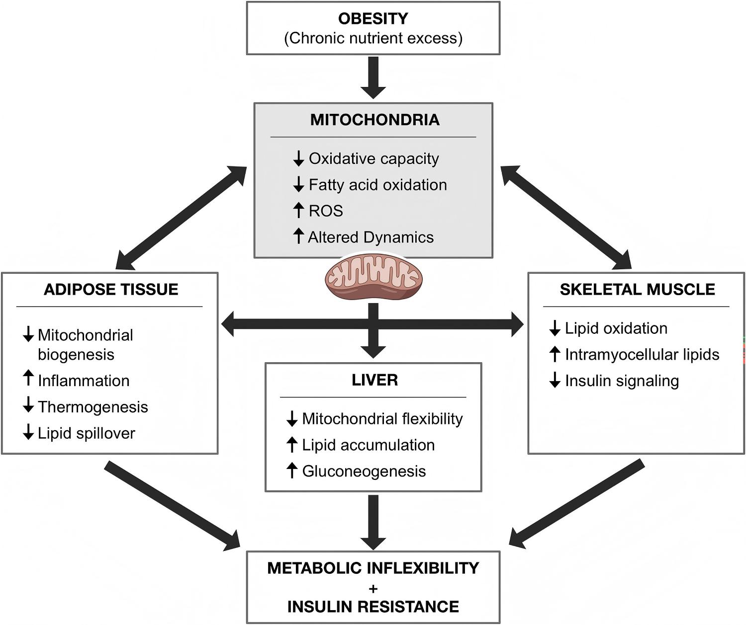 Fig.1 How obesity-driven mitochondrial damage triggers metabolic inflexibility and insulin resistance. (Neto, et al.; 2026)
Customer Review
Unbiased 3D Tissue Reconstruction and Morphometric Data for Regulatory Submissions
"We were struggling to generate clear, reproducible data showing how our lead small molecule impacted mitochondrial structure in high-fat diet mouse tissues. Protheragen took over our ex vivo tissue processing and delivered incredibly sharp, automated 3D network reconstructions. The quantification of mitochondrial branching and aspect ratios gave us the clear, unbiased data we needed for our upcoming regulatory submission. We are already mapping out our next live-cell kinetic project with their team."
Dr. A. T., Metabolic Therapeutics Startup
Automated High-Content Screening That Streamlines Lead Optimization in Lipotoxicity Models
"The automated image analysis from Protheragen eliminated the manual bottleneck in our screening workflow. Their team was able to clearly show a dose-dependent rescue of mitochondrial networks from fragmented puncta back to healthy networks in our lipid-loaded muscle cell lines. Their scientific expertise and deep understanding of lipotoxicity mechanisms made them a true extension of our pharmacology team."
Dr. E. V., Lead Optimization Division
Frequently Asked Questions
-
What types of cell and tissue samples can be analyzed using this service?
We regularly work with primary mammalian adipocytes, skeletal muscle cells (such as differentiated L6 or C2C12 cells), hepatocytes, and cardiotoxic cellular models. We can also process fresh or optimally frozen tissue sections from adipose tissue depots, liver, and skeletal muscle tissue collected during your in-house animal studies.
-
How does Protheragen handle high-fat or lipid-laden samples without ruining the fluorescent signal?
Large intracellular lipid droplets in mature adipocytes can sometimes cause light scattering or non-specific staining. We use specialized, validated permeabilization and blocking protocols along with highly photostable fluorophores to ensure sharp, high-contrast mitochondrial imaging even in tissues with heavy lipid accumulation.
-
Are your services appropriate for clinical trial patient sample monitoring?
No, our platform is built exclusively for preclinical research. We specialize in in vitro cell lines, primary cell cultures, and ex vivo animal tissue models, helping you secure strong mechanistic data before moving toward clinical development.
-
What specific morphometric parameters are included in your final data report?
Our standard reports include a comprehensive suite of data points: individual mitochondrial length, width, aspect ratio, perimeter, circularity, network branching frequency, total network length, and the ratio of fragmented vs. elongated mitochondria across all experimental groups.
-
Can we test the effects of small-molecule inhibitors of mitochondrial fission, like Mdivi-1 or P110?
Absolutely. We frequently use these specific inhibitors as positive controls in our assays to benchmark your candidate compounds and confirm if your molecules achieve similar or superior structural protection against lipotoxic fragmentation.
-
How do you measure live mitochondrial dynamics without causing phototoxic damage to the cells?
We use ultra-sensitive, high-speed resonance scanning confocal setups combined with low-intensity laser excitation and advanced digital deconvolution. This approach gives us excellent spatial resolution while keeping cells healthy during live-cell time-lapse experiments.
-
What is the typical turnaround time from sample receipt to the final data package?
Turnaround times generally range between 3 to 6 weeks, depending on the number of samples, the complexity of the cell models, and whether you require live-cell tracking or fixed tissue processing. We establish clear timelines during our initial project consultation.
-
Can this service help explain the mechanism behind elevated reactive oxygen species (ROS) in obese models?
Yes, because structural fragmentation and ROS production are closely linked. By pairing structural analysis with co-staining techniques, we can correlate specific structural changes (like fragmentation) with localized oxidative stress or mitochondrial membrane depolarization.
Contact Us
Sifting through the complex structural changes that occur during obesity-induced lipotoxicity requires highly specialized imaging tools and deep technical expertise. Protheragen provides a dedicated obesity-related mitochondrial morphology & structural analysis service that turns complex cellular architecture into reliable, quantitative datasets. From automated high-content structural profiling to direct kinetic measurements of fusion and fission, our platform gives your preclinical discovery team the precise data needed to advance promising metabolic candidates. Contact Protheragen now for more information and a personalized consultation.
Reference
- Neto, W.H.J.; et al. Mitochondrial remodeling in obesity: mechanistic links to impaired energy metabolism and therapeutic perspectives. Physiology & Behavior. 2026, 315, 115427. (CC BY 4.0)
All of our services and products are intended for preclinical research use only and cannot be used to diagnose, treat or manage patients.