
Obesity-Related Mitochondrial Fusion and Fission Assay Service
InquiryMitochondria do not just sit passively inside cells generating ATP; they form a highly dynamic, interconnected network that is constantly shifting through opposing processes known as fusion and fission. In healthy metabolic states, a delicate balance keeps this network tuned to the cell's energetic needs. However, chronic nutrient overload and metabolic stress drastically alter this landscape. Recent preclinical research reveals that obesity disrupts this equilibrium, frequently pushing the system toward hyper-fragmentation. This shift occurs when structural networks shatter into smaller, inefficient structural units, directly impacting the capacity to burn fat and maintain systemic insulin sensitivity.
Preclinical Mitochondrial Dynamics Services for Obesity Drug Discovery
Protheragen provides an optimized preclinical testing platform designed to evaluate these morphological transitions and structural variations in adipose, skeletal muscle, and hepatic cellular models. Our assays monitor the shifting balance between outer/inner membrane fusion and cytosolic fission machinery under simulated pathophysiological conditions. By quantifying these structural adjustments, we help drug development programs evaluate small-molecule therapeutics, biological candidates, and dietary compounds intended to restore structural stability and improve metabolic resilience.
Core Technologies
To accurately characterize structural dynamics within cell populations, we use a multivalent technical approach that combines high-resolution imaging with precise biochemical quantifications.
Automated, multi-channel laser scanning confocal systems provide detailed structural visualization of cellular networks. Specialized software segments the network to extract metrics including branch length, network complexity, aspect ratio, and fragmentation indices.
We monitor critical fusion markers—mitofusin 1 (Mfn1), mitofusin 2 (Mfn2), and optic atrophy 1 (Opa1)—alongside key fission components like dynamin-related protein 1 (Drp1) and fission 1 protein (Fis1). This tracking uses automated Western blotting and high-throughput qPCR.
Fission activation relies heavily on the post-translational modification of Drp1 and its subsequent translocation from the cytoplasm to the outer mitochondrial membrane. Our assays specifically quantify phosphorylation states (such as Ser616 and Ser637) using advanced cell fractionation procedures and targeted immunoassay platforms.
Service Scope
Our specialized assay pipeline covers a broad range of preclinical models and analytical markers to ensure deep insight into metabolic drug candidates.
- Cellular Model Customization
We handle a diverse selection of immortalized lines and primary cell cultures relevant to metabolic syndrome research:
- Adipocyte Frameworks: Differentiated 3T3-L1 cells and primary white/brown preadipocytes exposed to palmitate or oleate to model lipotoxicity.
- Myotube Systems: C2C12 myotubes and primary skeletal muscle cells used to examine the intersection of structural fragmentation and insulin signaling cascades.
- Hepatic Models: HepG2 cells and primary hepatocytes, focusing on steatosis development and its impact on lipid accumulation and structural breakdown.
- Quantitative Morphological Profiling
Instead of relying on subjective visual assessments, we convert complex network images into clear, actionable datasets. Our automated analysis pipelines measure:
- Fragmentation Index: The ratio of individual, isolated puncta to total network area, indicating severe network breakdown.
- Aspect Ratio and Form Factor: Geometric measurements that track the elongation and branching complexity of individual organelles.
- Network Interconnectivity: Quantitative tracking of junctions and nodes to determine how well the network maintains functional structural continuity.
- Molecular Regulatory Profiling
We analyze the expression and behavior of key regulatory proteins driving these structural shifts:
- GTPase Abundance Tracking: Evaluating changes in Mfn1, Mfn2, and Opa1 levels to determine if a compound supports network fusion.
- Fission Activation Analysis: Measuring total Drp1 expression alongside its specific phosphorylation changes to identify mechanisms that prevent excessive fragmentation.
- Subcellular Translocation Assays: Isolating cytosolic and membrane fractions to track the physical recruitment of fission components to the outer organelle membrane.
Connect with Our Preclinical Scientific Team to Receive a Detailed Technical Proposal.
Workflow
Translating a complex metabolic hypothesis into reliable, reproducible data requires a structured experimental framework that maintains rigorous quality controls at every milestone.
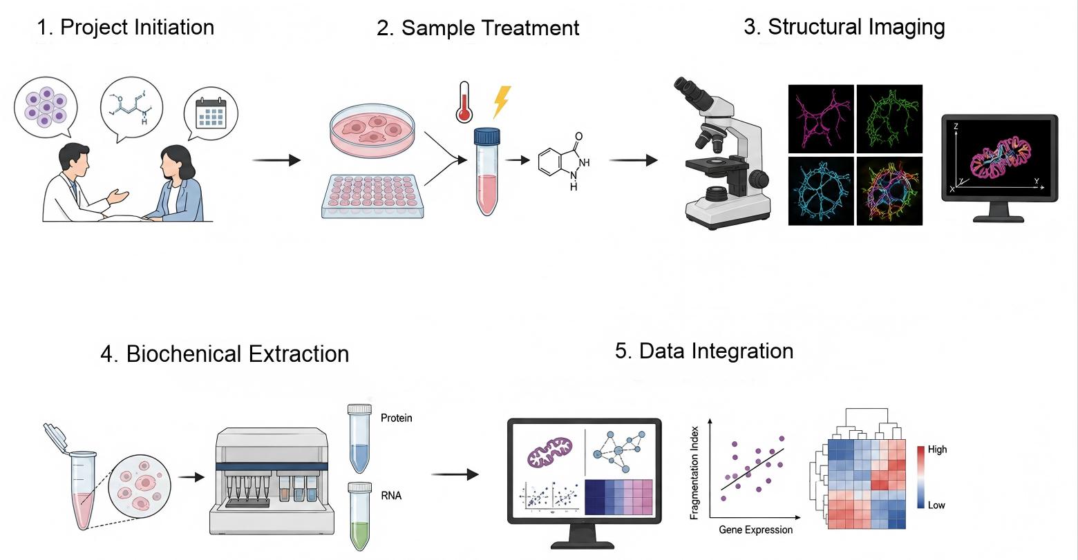
- Step 1: Detailed consultation defines your study parameters, specific cellular models, compound concentrations, and timeline requirements.
- Step 2: Preclinical cell lines or primary tissues undergo metabolic stress induction alongside compound incubation protocols.
- Step 3: High-resolution live-cell or fixed staining protocols capture complex multi-dimensional mitochondrial network architecture.
- Step 4: Cell lysates undergo automated protein and RNA extraction to prepare for downstream regulatory tracking.
- Step 5: Comprehensive statistical profiling correlates structural network fragmentation metrics directly with target regulatory expression.
Fields of Application
Because structural network transitions serve as a core indicator of metabolic wellness, this specialized testing platform provides critical value across multiple stages of early-stage discovery.
- Preclinical Lead Optimization: Screening small-molecule or biologic libraries to identify structures that prevent excessive fragmentation under high-lipid stress conditions.
- Mechanistic MoA Validation: Confirming whether metabolic drug candidates operate by activating fusion regulators or directly inhibiting fission machinery.
- Nutraceutical Screening: Evaluating natural extracts, fatty acid variants, and functional food ingredients for their ability to maintain structural integrity during caloric overload.
- Biomarker Discovery Support: Identifying downstream changes in protein expression or phosphorylation that correlate with restored network morphology, aiding future translation.
Advantages
Selecting the right research partner means finding a team that delivers more than just raw data; it requires deep expertise that converts complex biological images into clear, actionable mechanisms.
Validated Preclinical Disease Models
Our platform uses carefully optimized, physiologically relevant metabolic stress protocols. These structural models reliably mimic the network fragmentation and downstream insulin resistance pathways observed in diet-induced metabolic dysfunction. This alignment gives you confidence that your compound's performance in our assays reflects true biological mechanisms.
High-Content, Objective Quantification
We avoid qualitative visual scoring by utilizing automated segmentation algorithms. This ensures highly reproducible datasets with low coefficient of variation (CV) values, making it easier to detect subtle, dose-dependent improvements in network architecture.
Mechanistic Depth and Assay Synergy
Rather than simply reporting whether a network is fragmented, we connect structural alterations directly to specific protein modifications and transcriptional regulators. This multi-layered data confirms your candidate's exact molecular mode of action, providing essential evidence for pipeline progression.
Connect With Our Scientific Experts Today to Design a Tailored Testing Protocol.
Customer Review
Validating Mechanism of Action and Dose-Dependent Network Restoration in Myotube Models
"We needed to confirm the exact mechanism of action for our lead metabolic candidate, specifically regarding how it influenced organelle structure in lipid-loaded muscle models. Protheragen provided high-content imaging datasets paired with clear protein expression metrics that clearly demonstrated a dose-dependent reduction in fragmentation. The quality of the data and clarity of the report helped us make confident pipeline decisions. We plan to partner with them again for our upcoming structural profiling studies."
Dr. P. B., Cardiovascular & Metabolic Therapeutics
High-Throughput Screening of Natural Compounds Using Optimized Adipocyte Frameworks
"Our team was looking for a reliable partner to screen natural extracts for their ability to preserve network structure under high-nutrient stress. The technical team at Protheragen adjusted their adipocyte assays to fit our specific screening requirements perfectly. Their automated morphology metrics provided clean, reproducible data that allowed us to identify two key focus compounds. Their constant communication and expertise made the entire collaboration highly productive."
Dr. P. M., Metabolic Health & Nutrition Lead
Frequently Asked Questions
-
Can your platform distinguish between direct fission inhibitors and indirect metabolic adaptors?
Yes, we can. By combining high-content structural imaging with subcellular fractionation assays that track Drp1 membrane recruitment, we differentiate between direct structural targeting and broader upstream metabolic improvements.
-
What cell models do you recommend for studying obesity-driven skeletal muscle resistance?
We generally suggest using differentiated C2C12 myotubes or primary skeletal muscle cells exposed to high palmitic acid concentrations, as this model reliably induces fragmentation and impairs insulin signaling pathways.
-
How do you ensure your automated imaging tools don't count background artifacts as fragmented organelles?
Our imaging process utilizes optimized multi-channel staining alongside strict size, intensity, and shape thresholds during automated segmentation, effectively filtering out non-specific background signals.
-
Can we supply our own proprietary cell lines or specific transgenic models for these studies?
Absolutely. We frequently onboard client-provided cell lines or specific primary tissues. Our technical team will work with you to adapt our staining and isolation protocols to your specific models.
-
Do you offer clinical testing services or analysis on human patient biopsy samples?
No, Protheragen focuses exclusively on preclinical research services. We do not provide clinical diagnostics or analyze samples from human clinical trials.
-
How do you maintain sample stability during protein extraction for phosphorylation analyses?
We use optimized lysis buffers supplemented with comprehensive protease and phosphatase inhibitor cocktails, maintaining strict temperature controls throughout extraction to safeguard fragile post-translational marks.
-
Is it possible to combine this structural assay with oxygen consumption rate (OCR) measurements?
Yes, pairing structural analysis with metabolic flux measurements provides a clearer view of how structural dynamics influence overall bioenergetic efficiency. Please mention this interest when discussing your project.
-
What control compounds do you typically include in these fragmentation assays?
We commonly use established reference molecules like Mdivi-1 to inhibit fission pathways, or vehicle controls under high-fat conditions to establish a baseline for maximum network fragmentation.
Contact Us
Maintaining a balanced mitochondrial network is vital for metabolic health, and understanding these structural dynamics is key to developing effective therapies for obesity-related conditions. Protheragen offers the validated models, high-content imaging systems, and molecular assays required to thoroughly evaluate your preclinical candidates. Contact Protheragen for more information on tailored solutions for your research.
All of our services and products are intended for preclinical research use only and cannot be used to diagnose, treat or manage patients.