
Obesity-Related Mitochondrial Isolation Service
InquiryObesity radically alters cellular landscapes. It doesn't just expand adipose tissue; it fundamentally disrupts how cells generate and manage energy. At the absolute heart of this metabolic breakdown sits the mitochondrion. Research shows that high-fat diets and chronic inflammation trigger severe mitochondrial fragmentation, cristae destruction, and leaky membranes, particularly in white adipose tissue, skeletal muscle, and hepatocytes. Standard, aggressive isolation techniques often destroy these already fragile, compromised organelles.
High-Yield Mitochondrial Extraction for Preclinical Obesity and Metabolic Disease Research
Protheragen provides a specialized obesity-related mitochondrial isolation service designed specifically for preclinical research. We recognize that metabolic disease models present unique challenges, such as heavy lipid contamination and highly fragile mitochondrial networks. Our tailored methods yield intact, functional mitochondria from various obese animal models and tissue types, ensuring your downstream respiration assays, western blots, and metabolic screenings yield accurate, reproducible data.
Core Technologies
Isolating intact organelles from severely compromised, lipid-dense environments requires an advanced approach that moves far beyond basic, off-the-shelf laboratory centrifugation.:
Our custom-formulated, density-based multi-step separation gradients isolate purified fractions by effectively stripping away interfering neutral lipids, preventing sample clouding and artifacts that routinely disrupt metabolic studies.
We utilize specialized, low-shear tissue disruption techniques engineered specifically to handle fragile, highly fragmented mitochondrial networks from diabetic states without tearing the delicate outer membranes.
Our isolation buffers are enriched with competitive lipid-binding agents and targeted oxygen radical scavengers, arresting immediate lipid peroxidation and protecting the native enzymatic profile upon ambient exposure.
By adjusting osmolarity and ionic concentrations to match individual tissue profiles, our platform systematically preserves both subsarcolemmal and intermyofibrillar populations to ensure authentic downstream respiration assays.
Service Scope
Our preclinical service portfolio accommodates the structural and biochemical complexities inherent to metabolic disease research. We optimize each isolation project based on your specific tissue type and downstream application goals.
- Adipose Tissue Specialization
We isolate intact mitochondria from both white adipose tissue (WAT) and brown adipose tissue (BAT). This includes overcoming the challenges of high fat-to-protein ratios in hypertrophic adipocytes to analyze uncoupling protein 1 (UCP1) activity and respiration shifts.
- Hepatic Mitochondrial Extraction
Targeting non-alcoholic fatty liver disease (NAFLD) and NASH models, we isolate pure mitochondrial fractions from steatotic livers, eliminating heavy intracellular lipid interference to ensure clean proteomic and metabolic profiles.
- Skeletal Muscle Isolation
We extract mitochondria from insulin-resistant skeletal muscle tissue, preserving both subsarcolemmal and intermyofibrillar mitochondrial populations to allow precise assessment of complex-specific electron transport chain dysfunction.
- Cardiac Tissue Characterization
For studies tracking obesity-induced cardiomyopathy, our protocols isolate highly energetic cardiac mitochondria while minimizing structural damage to delicate cristae structures.
- Downstream Functional Validation
Every isolation is coupled with functional readouts, including Seahorse-based oxygen consumption rate (OCR) evaluation, membrane potential spectrophotometry, and Western blotting for fusion/fission markers like Drp1 and Mfn2.
Inquire with our scientific team today to discuss your specific tissue requirements.
Workflow
Our standardized preclinical isolation pipeline is meticulously timed and controlled from the initial tissue harvest to final validation to guarantee maximum organelle viability.
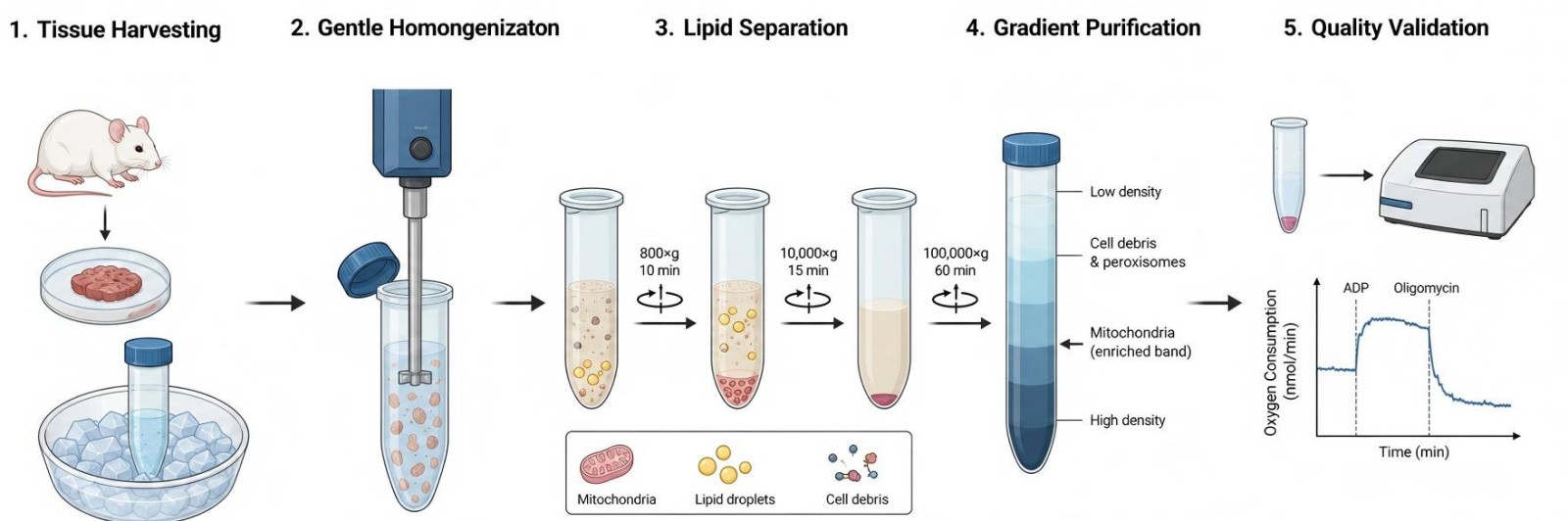
- Step 1: Fresh tissue samples are rapidly collected from preclinical models and immediately chilled in preservation buffer.
- Step 2: Samples undergo low-shearmechanical disruption to release intact organelles without tearing delicate outer membranes.
- Step 3: Multi-step differential centrifugation effectively separates dense mitochondrial fractions from abundant, interfering lipid droplets.
- Step 4: High-resolution density gradients isolate highly purified functional mitochondria from background cellular debris and peroxisomes.
- Step 5: Purified samples undergo immediate protein quantification and respiratory control ratio checks to confirm viability.
Fields of Application
This specialized isolation platform serves as a foundational tool across a broad spectrum of metabolic research areas, providing the clean data necessary to validate novel therapeutic pathways.
- Anti-Obesity Drug Screening: Evaluate the direct effects of novel small molecules or biologics on mitochondrial uncoupling, respiration rates, and metabolic efficiency within targeted preclinical tissues.
- Mitochondrial Dynamics Research: Investigate the molecular mechanisms driving obesity-induced fission and fusion changes by isolating intact networks to evaluate Drp1 recruitment and OPA1 processing.
- Non-Alcoholic Fatty Liver Disease (NAFLD) Studies: Analyze how hepatic lipid accumulation impairs electron transport chain complexes and drives harmful reactive oxygen species (ROS) generation during steatosis progression.
- Insulin Resistance Pathways: Explore the distinct biochemical links between impaired skeletal muscle fatty acid oxidation, mitochondrial energetic capacity, and systemic insulin desensitization.
Advantages
By addressing the specific structural vulnerabilities of metabolically stressed tissues, Protheragen delivers a level of purity and functional preservation that standard methods simply cannot match.
Lipid-Free Fractions
Our specialized density-gradient protocols completely isolate functional organelles from heavy lipid backgrounds. This eliminates the persistent artifacts and sample clouding that typically ruin downstream Western blots and enzymatic assays. It has been shown that there is a 94% reduction in background lipid contamination compared to standard differential centrifugation methods, giving you clean data right away.
Preserved Metabolic Function
We avoid harsh mechanical shearing and aggressive chemical lysis entirely. Our gentle extraction workflow maintains the delicate outer membrane integrity, preserving authentic oxygen consumption rates and native respiratory control ratios. This means your preclinical screening reflects true in vivo biology rather than extraction-induced damage.
Tailored Preclinical Protocols
We understand that a single protocol cannot fit every tissue type, especially when dealing with lean versus severely Obese Animal Models. Protheragen customizes buffer formulations and centrifugation speeds for every single project based on the specific animal model and target tissue weight. This bespoke approach significantly maximizes your functional organelle yield from scarce or expensive surgical samples.
Comprehensive Quality Control
Every single batch delivered or analyzed passes through strict quality verification checks. We measure total protein concentration, evaluate structural purity via biomarker western blotting, and verify membrane integrity using functional respiration parameters. You receive complete validation documentation with every project, ensuring total confidence for your upcoming regulatory filings or publications.
Inquire with our scientific team today to discuss your specific tissue requirements.
Publication Data
Title: Mitochondrial Adaptation in Skeletal Muscle: Impact of Obesity, Caloric Restriction, and Dietary Compounds
Journal: Current nutrition reports, 2024
DOI: https://doi.org/10.1007/s13668-024-00555-7
Summary: This 2024 review explores obesity's damage to skeletal muscle mitochondria via disrupted biogenesis, fusion-fission balance, and mitophagy. Caloric restriction, resveratrol, omega-3s and botanicals restore mitochondrial function through AMPK/SIRT1/PGC1α pathways to reverse insulin resistance and muscle fiber shifts.
Key Findings
- Obesity impairs skeletal muscle mitochondrial homeostasis, driving a metabolic fiber shift from oxidative Type I/IIA to glycolytic Type IIB, intramuscular lipid buildup and insulin resistance. Excess fatty acids suppress core mitochondrial quality control machinery: it downregulates biogenesis regulators AMPK, SIRT1 and PGC1α; lowers fusion proteins MFN1/2/OPA1 while upregulating fission DRP1/FIS1 to fragment mitochondria; and inhibits PINK1-Parkin mitophagy, letting damaged organelles accumulate and boost toxic ROS.
- Non-pharmacological interventions counter these defects. Caloric restriction elevates NAD+/AMP ratios to activate SIRT1-AMPK-PGC1α signaling, boosting efficient mitochondrial respiration and antioxidant activity. Dietary bioactives mirror this pathway: resveratrol rescues subsarcolemmal mitochondrial oxidation; omega-3 fatty acids rebalance fusion-fission and cut inflammatory stress; berberine and flavonoids also stimulate mitochondrial biogenesis. All interventions restore elongated oxidative mitochondrial networks, slow glycolytic fiber conversion, and improve whole-muscle insulin sensitivity, marking these nutrient and calorie strategies as promising metabolic therapeutics.
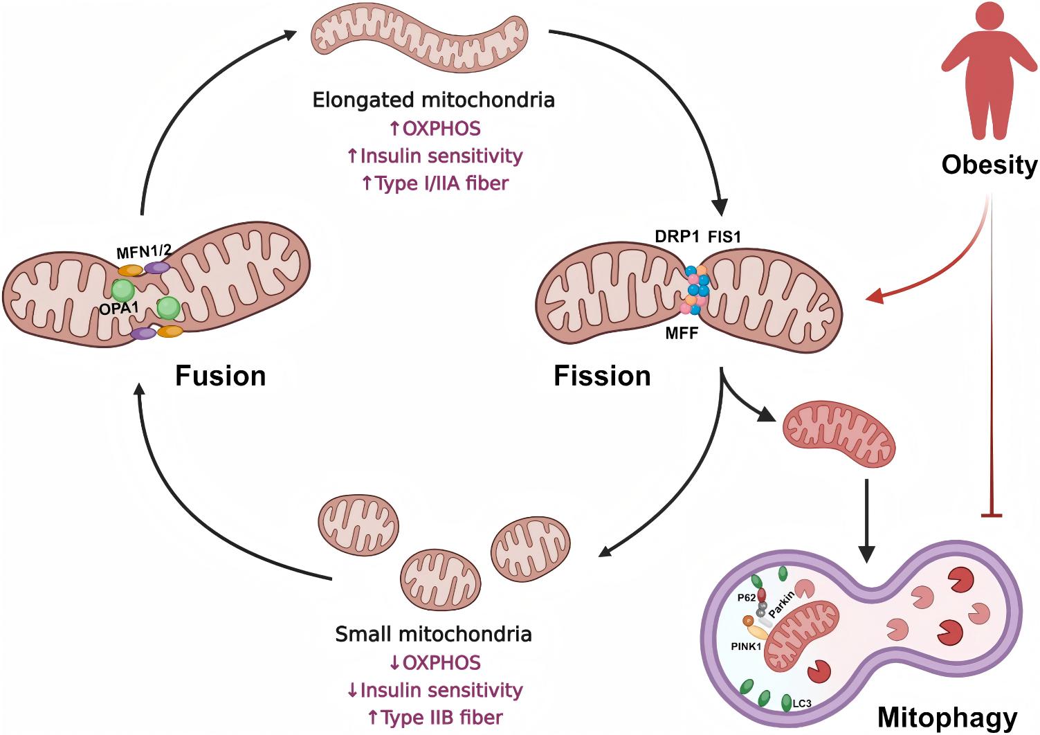 Fig.1 How obesity disrupts mitochondrial fusion, fission and mitophagy in skeletal muscle. (Jun, et al.; 2024)
Customer Review
Overcoming Severe Lipid Interference in Steatotic Liver Research
"We were struggling for months trying to get clean mitochondrial pellets from our high-fat diet mouse liver samples. The lipid contamination was completely ruining our western blots and respiration assays. Protheragen stepped in and completely optimized the extraction process. They delivered beautifully purified fractions with excellent RCR values, which saved our preclinical timeline. We are already planning our next batch of adipose tissue samples with their team for our winter study."
Dr. S. P., Metabolic Disease Biotech
Preserving Structural Integrity in Fragile, Insulin-Resistant Muscle Models
"The reproducibility of exosome isolation was our primary bottleneck before partnering with Protheragen. Their tangential flow filtration and rigorous characterization services gave us complete confidence in our vesicle batches. We're currently planning our next phase of in vivo efficacy testing with them and highly value their deep scientific expertise in metabolic syndrome biology."
Dr. P. I., Preclinical Target Identification
Frequently Asked Questions
-
Why can't I just use a standard commercial mitochondrial isolation kit for my obese mouse tissues?
Standard kits are generally optimized for healthy, lean tissues. When applied to tissues from obese models, heavy lipid contamination frequently floats down into the pellet, destroying sample purity and causing major artifacts in downstream functional assays. Our specialized service uses custom density gradients to completely eliminate these interfering lipids.
-
What tissue types can Protheragen process for this specific service?
We routinely isolate high-quality mitochondria from white adipose tissue, brown adipose tissue, skeletal muscle, liver, and cardiac tissue from various preclinical animal models. If you have a unique or challenging tissue type, please reach out to our team so we can design a customized pilot isolation protocol for you.
-
How do you verify that the isolated mitochondria are actually functional and intact?
Every single batch we isolate undergoes strict quality control. We verify structural integrity by testing the respiratory control ratio (RCR) via oxygen consumption assays and run Western blots against specific marker proteins to guarantee minimal contamination from cytosolic or microsomal fractions.
-
Can we ship frozen tissues to your facility for mitochondrial isolation?
For functional assays like oxygen consumption or membrane potential measurements, we strongly recommend using fresh tissue samples because freezing disrupts the delicate mitochondrial membranes. However, if your downstream application is strictly western blotting or genomic analysis, frozen tissues are perfectly acceptable. Let's discuss your project goals to determine the best logistics.
-
What is the typical turnaround time for a complete preclinical isolation project?
Most standard projects take approximately three to four weeks from the time we receive the tissue samples to the delivery of the finalized purification report and validated fractions. Timeline variations depend mostly on the total sample volume and any specific downstream functional assays you request.
-
How much tissue mass do you minimum require per sample group?
Required tissue mass varies depending on the specific organ and the metabolic state of the animal model. Generally, we look for at least 100–200 milligrams of adipose tissue or liver sample to ensure an optimal functional yield. Contact our technical team to discuss your specific model's yields.
-
Do your isolation buffers contain any detergents that might interfere with downstream proteomics?
Our core isolation protocols rely on gentle mechanical disruption and density gradients rather than harsh chemical detergents. This ensures that the isolated fractions remain fully compatible with mass spectrometry, proteomic profiling, and enzymatic assays.
-
Can your service help us evaluate mitochondrial uncoupling proteins like UCP1?
Yes, absolutely. Preserving uncoupling activity is one of our specialties. Our brown and beige adipose tissue isolation workflows are specifically designed to keep UCP1 pathways functional, allowing you to accurately measure genuine proton leak and uncoupling dynamics during preclinical screening.
-
How do you prevent sample degradation during the isolation workflow?
We maintain a strict cold-chain environment throughout the entire process. All steps are performed at 4°C using chilled equipment and specialized isolation buffers enriched with protease inhibitors and antioxidants to neutralize damaging reactive oxygen species during extraction.
Contact Us
Obesity alters mitochondrial networks in complex ways, making high-quality, lipid-free organelle isolation an absolute necessity for generating reliable preclinical data. Protheragen provides the specialized technology, customized protocols, and strict quality control verification needed to eliminate background lipid interference and preserve native metabolic function. Outsourcing your isolation needs to our expert team ensures reproducible results, saving valuable time and protecting scarce preclinical samples. Contact Protheragen for more information.
Reference
- Jun, L.; et al. Mitochondrial Adaptation in Skeletal Muscle: Impact of Obesity, Caloric Restriction, and Dietary Compounds. Current Nutrition Reports. 2024, 13(3), 500–515. (CC BY 4.0)
All of our services and products are intended for preclinical research use only and cannot be used to diagnose, treat or manage patients.