
Brain Slice Patch Clamp Service
InquiryUnderstanding the central control of energy balance requires direct measurement of neuronal excitability and synaptic transmission in homeostatic brain centers. Hypothalamic circuits—particularly neurons within the arcuate nucleus (ARC), ventromedial hypothalamus (VMH), lateral hypothalamus (LH), and brainstem regions like the nucleus tractus solitarius (NTS)—dynamically process metabolic signals such as leptin, insulin, ghrelin, and nutrient metabolites.
Precision Brain Slice Patch Clamp Services for Metabolic and Obesity Research
Protheragen offers specialized electrophysiology services designed to decode these complex neural networks. Our ex vivo brain slice patch-clamp assay platform gives researchers high-resolution insight into ion channel kinetics, spontaneous and evoked synaptic activity, and membrane potential responses under specific metabolic, pharmacological, or dietary conditions. By evaluating functional neural activity in rodent models of diet-induced obesity (DIO) or genetic metabolic disease, our preclinical services bridge the gap between gene or molecular target discovery and systemic therapeutic efficacy.
Core Technologies
Our electrophysiology suite leverages complementary manual and automated patch clamp platforms to address the specific functional demands of obesity research:
Enables high-fidelity measurements of single-cell action potential firing, resting membrane potential, input resistance, and active ion currents (voltage-gated Na+, K+, and Ca2+ currents) in targeted hypothalamic and hindbrain subpopulations.
Quantifies miniature, spontaneous, and evoked inhibitory/excitatory postsynaptic currents (mIPSCs/mEPSCs, sIPSCs/sEPSCs, eIPSCs/eEPSCs) to determine whether metabolic signals act presynaptically or postsynaptically.
Integrates transgenic reporter lines (e.g., POMC-GFP) with infrared differential interference contrast (IR-DIC) and fluorescence microscopy to reliably patch specific neurochemical lineages.
Employs rapid solution exchange setups to test real-time neuronal responses to peptide hormones, nutrient molecules, candidate small molecules, or biologic therapeutics at physiological temperatures (32-34°C).
Service Scope
Protheragen provides comprehensive ex vivo electrophysiology assays focused on neuroendocrine control of feeding behavior and systemic metabolism:
Arcuate Nucleus Circuit Profiling
- Pro-opiomelanocortin (POMC) Neurons
Quantification of intrinsic excitability, tonic firing rates, and synaptic inputs in response to anorexigenic compounds, leptin, or GLP-1 receptor agonists.
- Agouti-Related Peptide (AgRP) / Neuropeptide Y (NPY) Neurons
Assessment of hyperpolarization or synaptic suppression induced by orexigenic or anorexigenic ligands in lean versus DIO animals.
- Presynaptic vs. Postsynaptic Dissection
Paired-pulse ratio (PPR) analysis and miniature EPSC/IPSC recordings in the presence of tetrodotoxin (TTX) to pinpoint the exact site of action.
Paraventricular & Lateral Hypothalamic Pathways
- PVH Single-Cell Recording
Evaluation of oxytocin-, CRH-, or MC4R-expressing neurons receiving inputs from ARC projection sites under acute drug application.
- LH Glucose-Sensing Subpopulations
Assessment of orexin/hypocretin and MCH neuron responses to ambient glucose fluctuations and metabolic peptide challenges.
Hindbrain Energy Balance Circuits
- NTS & Dorsal Motor Nucleus of the Vagus (DMV)
Profiling gut-brain axis signal integration, including vagal afferent inputs and responses to CCK, PYY, or dual/triple incretin mimetics.
Custom Metabolic State Profiling
- Diet-Induced Obesity (DIO) vs. Lean Comparisons
Profiling slice electrophysiology across high-fat diet (HFD) fed rodents and age-matched chow-fed controls to evaluate leptin/insulin resistance at the single-cell level.
- Genetically Modified Mouse Models
Validation of gene knockouts, knock-ins, or viral vector (AAV-Cre/DIO) manipulations directly on target cell electrophysiology.
Connect with our scientific specialists today.
Workflow
From careful tissue harvesting and high-viability slicing to targeted whole-cell recording and thorough data analysis, our systematic workflow ensures clean, reliable physiological data at every stage.
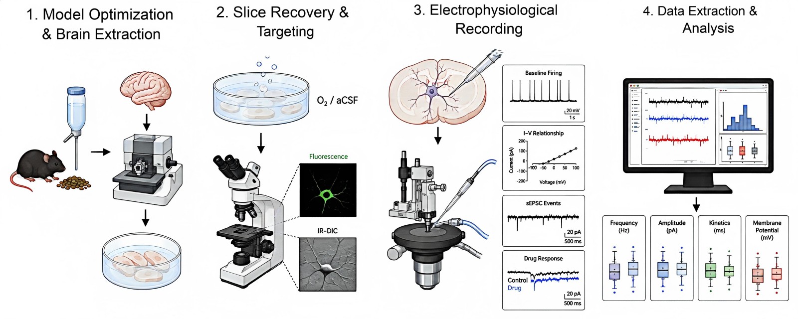
- Step1: Animals undergo specific dietary regimens before humane brain extraction and ultra-precise vibratome sectioning into protective physiological recovery buffers.
- Step2: Brain slices recover in oxygenated artificial cerebrospinal fluid (aCSF) before targeted fluorescent and IR-DIC identification of specific neurons.
- Step3: Stable whole-cell or cell-attached configurations measure baseline firing, current-voltage relationships, synaptic events, and acute drug responses.
- Step4: Signal traces undergo automated and manual filtering to quantify frequency, amplitude, kinetics, and membrane potential shifts across cohorts.
Fields of Application
From early-stage target validation to mechanistic off-target screening, our specialized slice electrophysiology platform empowers research across diverse pipelines in central metabolic and neuroendocrine drug discovery.
- Preclinical Anti-Obesity Drug Discovery: Screening novel GLP-1, GIP, glucagon, or amylin receptor co-agonists for direct central nervous system activity and neuronal modulation.
- Central Metabolic Safety & Off-Target Profiling: Evaluating whether systemic candidates cause unintended activation or suppression of off-target hypothalamic or autonomic neural circuits.
- Mechanistic Target Validation: Confirming the functional cellular mechanism of novel G-protein coupled receptors (GPCRs), ion channels, or transporters implicated in body weight regulation.
- Leptin and Insulin Resistance Research: Investigating the cellular mechanisms of impaired neuronal responsiveness during progressive high-fat diet feeding regimens.
Advantages
Our brain slice patch clamp service combines deep neuroendocrine expertise with rigorous quality control standards to provide ultra-precise, publication-ready electrophysiological data for your lead candidates.
Unrivaled Hypothalamic Expertise
Our electrophysiologists specialize exclusively in delicate hypothalamic and brainstem preparations, maintaining exceptional slice viability and cell-yield consistency even when working with aged or obese animal tissue. It is confirmed that our prep stability holds over extended recording windows.
Fluorescence-Guided Cell Identification
We routinely combine reporter mouse models with high-resolution optics, guaranteeing that recordings are collected strictly from verified cell types such as POMC, AgRP, or MC4R neurons rather than mixed surrounding populations.
Physiologically Relevant Pharmacological Assays
By implementing rapid perfusion systems with tight thermal regulation, we deliver reproducible dose-response curves for peptide drugs and Small Molecules under physiological conditions.
Fully Customized Preclinical Study Designs
We tailor animal housing diets, age-matched cohorts, treatment durations, and acute slice perfusion protocols directly to your drug discovery milestones, delivering publication-ready dataset packages with complete raw traces.
Explore Options for Your Metabolic Project - Request a Consultation Today.
Publication Data
Title: Energy imbalance alters Ca2+ handling and excitability of POMC neurons
Journal: eLife, 2017
DOI: https://doi.org/10.7554/eLife.25641
Summary: This eLife study finds high-fat diet-induced obesity disrupts calcium handling in hypothalamic satiety POMC neurons: elevated resting cytosolic Ca2+, impaired mitochondrial Ca2+ uptake, reduced excitability via SK channel activation, linking mitochondrial dysfunction to blunted satiety signaling.
Key Findings
- DIO suppresses POMC neuron excitability (cell-intrinsic, not synaptic)
High-fat diet-induced obesity (DIO) hyperpolarizes POMC resting membrane potential, cuts spontaneous action potential frequency, and doubles silent non-firing neurons. Spike frequency adaptation (SFA) is stronger in obese POMC cells; GABA receptor blockers fail to reverse these changes, proving intrinsic neuronal defects drive hypoexcitability. - DIO disrupts global intracellular Ca2+ homeostasis in POMC neurons
Fura-2 imaging shows resting free cytosolic Ca2+is drastically elevated in DIO mice. Two core Ca2+ regulatory systems fail: endogenous cytosolic Ca2+ buffer capacity drops sharply, and plasma membrane Ca2+ extrusion rates slow significantly. - Mitochondrial Ca2+ storage capacity is impaired in obese POMC neurons
FCCP mitochondrial Ca2+ release assays reveal smaller Ca2+ efflux from DIO mitochondria, confirming reduced mitochondrial Ca2+ uptake/storage—consistent with prior MFN2 downregulation and disrupted ER-mitochondria contacts in obesity. - Intracellular Ca2+ levels directly control POMC firing behavior
Experimentally raising extracellular Ca2+ mimics DIO's blunted excitability; lowering extracellular Ca2+ restores depolarization, spontaneous spiking, and normalizes exaggerated SFA in silent obese POMC neurons. - SK Ca2+-activated K⁺ channels link high Ca2+ to neuronal silencing
The SK channel blocker apamin depolarizes silent DIO POMC neurons, restores action potential firing, reduces SFA, and lowers stimulation threshold currents. BK channel blockers produce no rescue effect, identifying SK channels as the key downstream mediator of obesity-induced hypoexcitability. - Novel primary conclusion
This study provides the first direct evidence that chronic high-calorie feeding causes progressive, detrimental breakdown of Ca2+ homeostasis in satiety-signaling POMC neurons, crippling their ability to mediate satiety signals during obesity development.
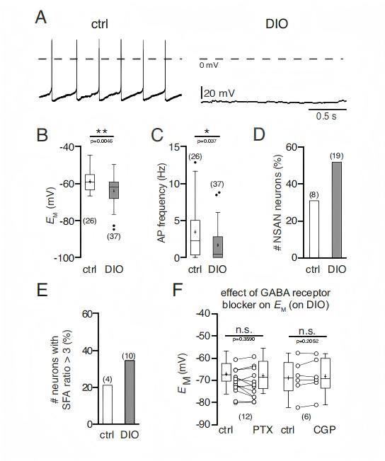 Fig.1 High-fat diet obesity suppresses spontaneous firing and hyperpolarizes satiety POMC hypothalamic neurons. (Lars, et al.; 2017)
Customer Review
Validation of Central Melanocortin Pathway Activity in DIO Models
"Working with Protheragen on our central melanocortin pathway project was a game changer. Their electrophysiology team successfully targeted POMC neurons in our high-fat diet animal cohort with remarkable consistency. The high-quality whole-cell recordings and clear synaptic data gave us the exact mechanistic proof of principle we needed for our internal decision gate. We are already setting up our next round of study protocols with them."
Dr. S. D., Biotechnology Partner
Elucidating Presynaptic vs. Postsynaptic Drug Mechanisms in Hindbrain Circuits
"We needed high-resolution slice patch-clamp recordings to distinguish presynaptic versus postsynaptic actions of our lead peptide drug in the NTS. Protheragen delivered flawless data with clear raw trace examples and robust statistical analyses within our tight timeline. Their communication throughout the project was top-notch, and we consider them an essential extension of our neuro-metabolic discovery platform."
Dr. P. S., Neuro-Metabolic Therapeutics
Frequently Asked Questions
-
What animal models do you typically use for brain slice patch-clamp assays in obesity research?
We routinely work with C57BL/6J mice subjected to diet-induced obesity (DIO) protocols, standard chow-fed controls, rat models, and transgenic reporter lines (such as POMC-eGFP or AgRP-Cre). We can also accommodate client-provided custom transgenic strains.
-
Can you record from specific cell types in the arcuate nucleus without fluorescent markers?
While transgenic fluorescent labeling is preferred for unambiguous identification of POMC or AgRP neurons, we can also target specific subregions based on cell morphology, anatomical landmarks, and post-recording RT-PCR or immunohistochemistry.
-
How do you maintain slice viability when working with tissues from severely obese animals?
Slicing tissue from old or high-fat diet-fed rodents is challenging due to increased lipid content and tissue fragility. We employ specialized protective recovery solutions, precise temperature control, and optimized slicing protocols to yield healthy, patchable neurons.
-
Can we test both acute drug responses and long-term pre-treatment effects?
Yes. We can perform acute bath applications of your compounds directly onto the slice during recording, or we can patch brain slices harvested from animals that underwent chronic in vivo dosing regimes.
-
What electrophysiological parameters are measured during standard compound screening?
Typical endpoints include resting membrane potential, spontaneous action potential firing frequency, input resistance, current-voltage (I-V) relationships, and parameters of miniature/spontaneous excitatory or inhibitory postsynaptic currents (mEPSCs, mIPSCs, sEPSCs, sIPSCs).
-
What is the typical turnaround time for a standard ex vivo patch-clamp study?
Depending on animal cohort availability and dietary pre-treatment timelines, standard recording studies are typically completed within 4 to 8 weeks, followed by comprehensive data analysis and reporting.
-
How do you differentiate between presynaptic and postsynaptic compound effects?
We utilize fast micro-perfusion of synaptic blockers (such as TTX to block action potentials, combined with receptor antagonists like picrotoxin or CNQX/AP5) alongside paired-pulse ratio protocol analysis to isolate presynaptic release probability from postsynaptic receptor sensitivity.
-
Can Protheragen handle custom viral vector expression validation in slice prep?
Absolutely. Clients frequently deliver viral constructs (AAV-DREADDs, optogenetic vectors, or shRNA) for stereotaxic brain microinjection at Protheragen prior to slice electrophysiology evaluation.
-
What format will the final data and deliverables take?
You receive a detailed scientific report featuring representative raw traces, quantified group comparisons (mean ± SEM with statistical testing), methodology descriptions, and complete raw data files formatted for submission or publication.
Contact Us
Protheragen is your trusted preclinical partner for unlocking the central neural control of metabolic disease. Our ex vivo brain slice patch-clamp services combine deep neuroendocrine domain expertise with cutting-edge electrophysiological platforms to advance your obesity research pipeline efficiently and reliably. Welcome to Contact Protheragen.
Reference
- Lars, Paeger.; et al. Energy imbalance alters Ca2+ handling and excitability of POMC neurons eLife. 2017, 6:e25641. (CC BY 4.0)
All of our services and products are intended for preclinical research use only and cannot be used to diagnose, treat or manage patients.