
Obesity-Related Mitochondrial Fluorescence Staining & Dynamics Service
InquiryObesity is far more than an accumulation of adipose tissue. At its heart, it represents a deep systemic disruption of energy balance that changes how cells function down to the organelle level. Mitochondria, the central powerhouses driving cellular respiration and lipid oxidation, often bear the brunt of this metabolic overload. When nutrient influx chronically outpaces cellular demand, the mitochondrial electron transport chain can falter, generating an excess of reactive oxygen species (ROS) and throwing off the delicate balance of mitochondrial fission and fusion. This structural and functional decline, frequently termed mitochondrial dysfunction, acts as a primary catalyst for downstream insulin resistance, chronic low-grade inflammation, and hepatic steatosis.
Visualizing Metabolic Rescue: Preclinical Mitochondrial Dynamics Services for Obesity Drug Discovery
To help therapeutic developers unpack these intricate mechanisms, Protheragen offers a specialized preclinical toolkit: our obesity-related mitochondrial fluorescence staining & dynamics service. Designed specifically for in vitro and ex vivo preclinical exploration, this platform allows researchers to visually map, track, and quantify real-time structural shifts, membrane potential alterations, and oxidative stress signatures within living cells. By examining these organelle-level dynamics in high-fat or lipotoxic culture environments, we help teams understand how potential anti-obesity therapeutics modify metabolic resilience before moving toward late-stage development.
Core Technologies
To accurately capture the subtle shifts in organelle behavior driven by lipotoxicity, our platform relies on an integrated suite of high-resolution optics and functional fluorophores.
Our testing facility utilizes advanced automated confocal platforms optimized for live-cell kinetic tracking. These systems capture structural transitions over extended periods without disrupting the delicate cellular environment.
We deploy specialized chemical probes like JC-1 to monitor inner mitochondrial membrane polarization. The system tracks the physical shift from red J-aggregates in healthy cells to green monomers in failing, depolarized organelles.
To isolate metabolic stress markers, we use localized fluorogenic sensors that target intra-mitochondrial superoxide generation. This approach ensures a clean separation between organelle-specific ROS signals and general cytoplasmic background noise.
Our platform converts visual fluorescence data into objective metrics. Custom imaging software automatically traces and measures thousands of individual mitochondrial fragments per well, calculating precise parameters like aspect ratio, branch length, and network junctions.
Service Scope
Our preclinical service portfolio covers a wide variety of assays designed to give a complete view of mitochondrial function or failure under obesity-mimicking conditions. We look closely at structural layout and operational health, providing critical evidence for early-stage pipeline validation.
- Mitochondrial Membrane Potential Assays
We provide precise measurements of inner membrane polarization across various cell types, including primary hepatocytes and skeletal muscle cells exposed to high lipid loads. This lets teams track whether an experimental compound prevents the drop in membrane potential commonly driven by fatty acid toxicity, or if it helps stabilize the cellular energy state.
- Structural Morphology and Network Dynamics
Mitochondria are incredibly dynamic, constantly shifting between fusion and fission. In obese tissue environments, this balance frequently tilts toward fission, resulting in fragmented, inefficient organelles. Our imaging platform tracks parameters like total mitochondrial volume, roundness, aspect ratio, and the complexity of the branching network, helping you evaluate whether your candidate compound successfully encourages mitochondrial fusion and network rebuilding.
- Organelle-Specific Oxidative Stress Profiling
Nutrient excess overwhelms the electron transport chain, causing a sharp spike in superoxide generation. By using specialized mitochondrial-targeted fluorescent probes, we isolate and measure intra-mitochondrial ROS independently from cytoplasmic signals. This gives you a clear window into how your therapeutic candidates alter local oxidative stress.
- Mitophagy and Organelle Turnover Tracking
Healthy cellular metabolism relies on the selective clearance of damaged mitochondria via mitophagy. Our service profiles this clearance process by tracking the co-localization of mitochondrial markers with lysosomal compartments. We help you determine whether your drug candidate restores healthy organelle turnover in models of lipid overload.
- Adipocyte Differentiation and Lipid Accumulation Mapping
We offer multiplexed imaging that combines mitochondrial staining with lipophilic dyes to simultaneously track lipid droplet accumulation and organelle distribution during adipogenesis. This approach makes it easier to observe how mitochondrial architecture adjusts as cells mature into lipid-laden adipocytes.
Connect with Our Preclinical Scientific Team to Receive a Detailed Technical Proposal.
Workflow
Our structured preclinical assay timeline ensures maximum reproducibility and data depth by guiding every compound through a rigorous, multi-stage validation framework.
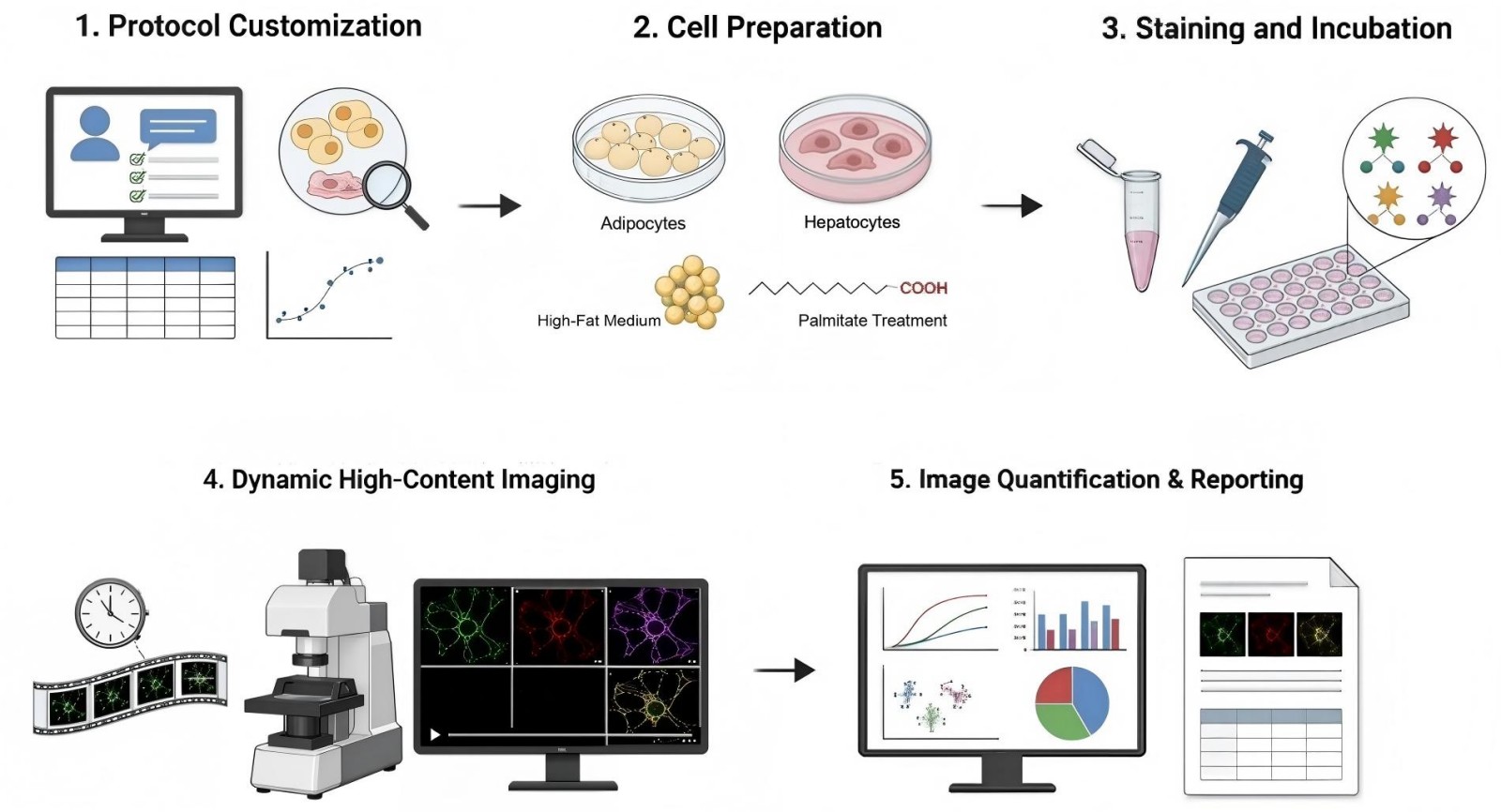
- Step 1: Collaborative alignment on cellular models, lipotoxic treatment criteria, and specific staining parameters tailored to project targets.
- Step 2: Culturing target adipocytes or hepatocytes under specialized high-fat or palmitate-induced metabolic stress conditions.
- Step 3: Applying precise, multiplexed fluorescent dye cocktails under carefully controlled, light-shielded environmental conditions for optimal binding.
- Step 4: Capturing real-time kinetic videos and multi-channel high-resolution confocal snapshots of the active mitochondrial networks.
- Step 5: Processing structural and intensity data through custom algorithms to deliver comprehensive, publication-ready metabolic reports.
Fields of Application
Our specialized staining and dynamics services support a variety of early-stage discovery initiatives:
- Anti-Obesity Small Molecule Screening: Evaluating library compounds to identify leads that preserve mitochondrial network integrity and prevent fragmentation under lipid stress.
- NASH/MASH Liver Disease Research: Tracking how fat accumulation in primary hepatocytes triggers mitochondrial calcium overload and structural collapse.
- Metabolic Inflexibility Studies: Analyzing how cells shift their organelle networks when moving between glucose and fatty acid utilization in high-fat culture models.
- Nutraceutical and Functional Food Testing: Validating the protective properties of natural extracts or dietary compounds against free-radical damage in metabolic cell lines.
Advantages
Partnering with Protheragen gives your discovery team access to an optimized testing environment designed to maximize data output while cutting down on experimental variance.
Advanced Multi-Channel Multiplexing Capabilities
Our imaging platforms allow for the simultaneous tracking of membrane potential, reactive oxygen species generation, and structural branching within the exact same cell sample. This deeply integrated testing setup extracts maximum data from every preclinical plate, reducing experimental variation and saving valuable compound volume.
Optimized Preclinical Models of Metabolic Strain
We have developed and validated specialized lipotoxicity protocols using primary cells and continuous lines conditioned with specific fatty acid complexes. This ensures our experimental environments closely mirror the intracellular lipid stress seen in in vivo disease states, making early discovery data much more relevant.
Custom Quantitative Image Segmentation Analysis
Instead of relying on basic visual checks, Protheragen utilizes automated image analysis pipelines that isolate and quantify thousands of individual mitochondrial fragments per well. We turn complex visual fluorescence data into clear numbers, mapping structural characteristics like aspect ratios and network junctions with zero human bias.
Proven Performance and Reproducible Outcomes
Our procedures are thoroughly optimized to minimize background fluorescence and prevent phototoxic damage during live-imaging sessions. Our automated assay frameworks maintain exceptional reproducibility, delivering Z'-factors consistently above 0.6, which provides the statistical rigor required for confident lead validation.
Submit Your Project Details via Our Secure Inquiry Portal
Customer Review
Validating Novel Compound Mechanism Under High-Fat Strain
"We needed to understand if our lead small molecule was directly protecting organelle structure or just acting as a general antioxidant under high-fat conditions. The imaging team at Protheragen provided beautifully clear, quantified data showing a real rescue of mitochondrial branching networks in our hepatocyte models. Their automated analysis gave our team the confidence we needed to move the program forward. We are already mapping out our next ex vivo project with them."
Dr. E. D., Biopharma Corporation
Seamless Workflow Integration and High-Data Reproducibility
"Working with Protheragen felt like an extension of our own internal lab group. Their scientists helped us refine our palmitate-stress protocols and delivered highly reproducible multi-channel fluorescence data that perfectly complemented our internal metabolic flux assays. The data package was clean, thorough, and ready for our internal reviews."
Dr. P. S., Metabolic Research Institute
Frequently Asked Questions
-
Can these assays be used to analyze human clinical samples?
No, our services are strictly limited to preclinical research. We operate entirely as an in vitro and ex vivo testing facility and do not process samples derived from human clinical trials or diagnostic pipelines.
-
What cell types can you run these obesity-mimicking studies on?
We regularly work with standard cell lines like 3T3-L1 adipocytes, HepG2 cells, and C2C12 myotubes, alongside primary rodent hepatocytes and adipose-derived stem cells. If you have a custom or proprietary cell model, we can adapt our staining protocols to fit your specific system.
-
How do you induce obesity-like conditions in an in vitro cell culture?
We typically introduce metabolic stress by exposing the cells to conjugated fatty acids, such as palmitate or oleate complexed with bovine serum albumin (BSA). This treatment successfully recreates the intracellular lipid accumulation and oxidative strain seen in metabolic disorders.
-
What is the difference between JC-1 and other mitochondrial stains you use?
JC-1 is uniquely useful for reading membrane potential because it shifts its emission color from green to red based on polarization. Other dyes we use are better suited for tracking long-term structural changes, where the signal remains stable regardless of minor fluctuations in membrane voltage.
-
Can you combine mitochondrial staining with other cellular readouts?
Yes, our system supports multi-channel imaging. We can easily pair mitochondrial dynamics tracking with additional stains for nuclear morphology, lipid droplet accumulation, or overall cell viability in a single assay run.
-
How do you prevent photobleaching and phototoxicity during live-cell imaging?
We use highly sensitive confocal systems and high-content imagers equipped with precise environmental controls. By optimizing exposure times and utilizing highly stable modern fluorophores, we keep light exposure low and preserve cell health throughout kinetic studies.
-
What kind of raw data will we receive at the end of the project?
We deliver a complete data package that includes high-resolution image files, analyzed statistical metrics (such as average branch lengths, network counts, and fluorescence ratios), and a thoroughly detailed summary report prepared by our lead analysts.
-
Are your image analysis workflows automated or manual?
Our entire segmentation process is automated. We utilize trained mathematical algorithms to identify and trace individual mitochondrial structures, which guarantees objective, highly reproducible datasets.
Contact Us
Mitochondrial breakdown underpins the progression of numerous metabolic disorders, making structural and functional organelle assays vital for modern drug discovery. Protheragen provides the high-resolution imaging infrastructure, validated lipotoxicity models, and automated quantitative analysis required to clearly evaluate your therapeutic assets in preclinical environments. Our specialized focus ensures your discovery team receives reliable, deep data to confidently guide your development decisions. Contact Protheragen for more information or to request a quote.
All of our services and products are intended for preclinical research use only and cannot be used to diagnose, treat or manage patients.