Decoding the Cellular Blueprint Linking Obesity and Metabolic Dysfunction to Pioneer Next-Generation Therapeutic Interventions
June 22, 2026

June 22, 2026
Visceral adipose tissue (VAT) is an immunologically active organ where protective tissue-resident macrophages (TRMs) maintain tissue homeostasis, while recruited monocyte-derived macrophages (MDMs) promote inflammation. Although MDM-driven inflammation during obesity is well-characterized, the survival mechanisms of protective TRMs during metabolic stress remain poorly understood.
Addressing this scientific gap, a study published in Nature Communications (2026) by Dutta et al., titled "Tissue-resident macrophage survival depends on mitochondrial function regulated by SerpinB2 in chronic inflammation," reveals how TRMs survive. Using preclinical models and human omental adipose tissue, the authors demonstrate that the intracellular protein SerpinB2 regulates mitochondrial function in TRMs, preserving this protective cell population to counter chronic inflammation and systemic insulin resistance.
The research team performed a series of highly detailed in vivo and in vitro experiments to understand the survival and anti-inflammatory mechanisms of TRMs.
Using CX3CR1-GFP/tdTomato fate-mapping and parabiosis, the authors confirmed that CX3CR1+ CCR2+ cells represent short-lived MDMs, while CX3CR1- CCR2- cells are long-term, self-renewing TRMs. RNA sequencing showed distinct transcriptomes: MDMs expressed pro-inflammatory genes linked to insulin resistance, whereas TRMs expressed insulin-sensitizing, anti-inflammatory markers. Correspondingly, obese human visceral adipose tissue (VAT) showed a marked loss of protective CCR2- TRMs alongside MDM accumulation.
In mice, high-fat diet (HFD) feeding triggered a progressive, apoptosis-driven decline in these protective TRMs, as evidenced by elevated active caspase-3 and annexin V. Notably, this decline is reversible; withdrawing the HFD restored the TRM pool via local proliferation (Ki-67+) rather than circulating monocyte recruitment.
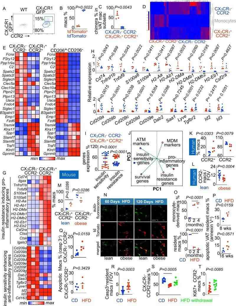 Fig.1 Resident and monocyte-derived macrophages in visceral fat show distinct immune and metabolic features during obesity progression. (Vasamsetti, et al., 2026)
Optogenetic depletion of VAT TRMs using BLE in LysM-Cre/ChR2 mice severely impaired glucose tolerance and insulin sensitivity, raising serum triglycerides (TG), free fatty acids (FFA), and free glycerol (FG). Depletion impaired insulin signaling (decreased pAkt/Glut4) and glucose-clearance gene expression while elevating pro-inflammatory cytokines.
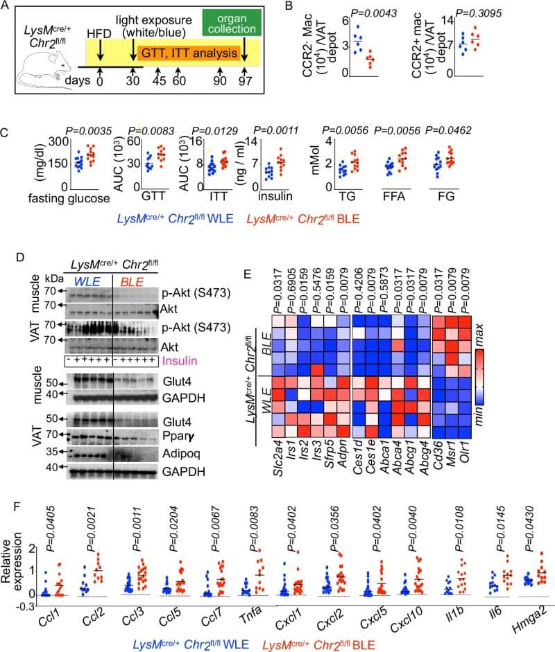 Fig.2 Loss of resident macrophages in visceral fat worsens inflammation and metabolic dysfunction in obese mice. (Vasamsetti, et al., 2026)
RNA sequencing identified SerpinB2 (encoding intracellular PAI-2) as highly and uniquely enriched in TRMs. Human TRMs displayed a similar enrichment. However, obesity significantly downregulated SerpinB2 in both mouse and human TRMs, with expression negatively correlating with body mass index (BMI). Palmitate treatment similarly suppressed SerpinB2 in bone marrow-derived macrophages (BMDMs).
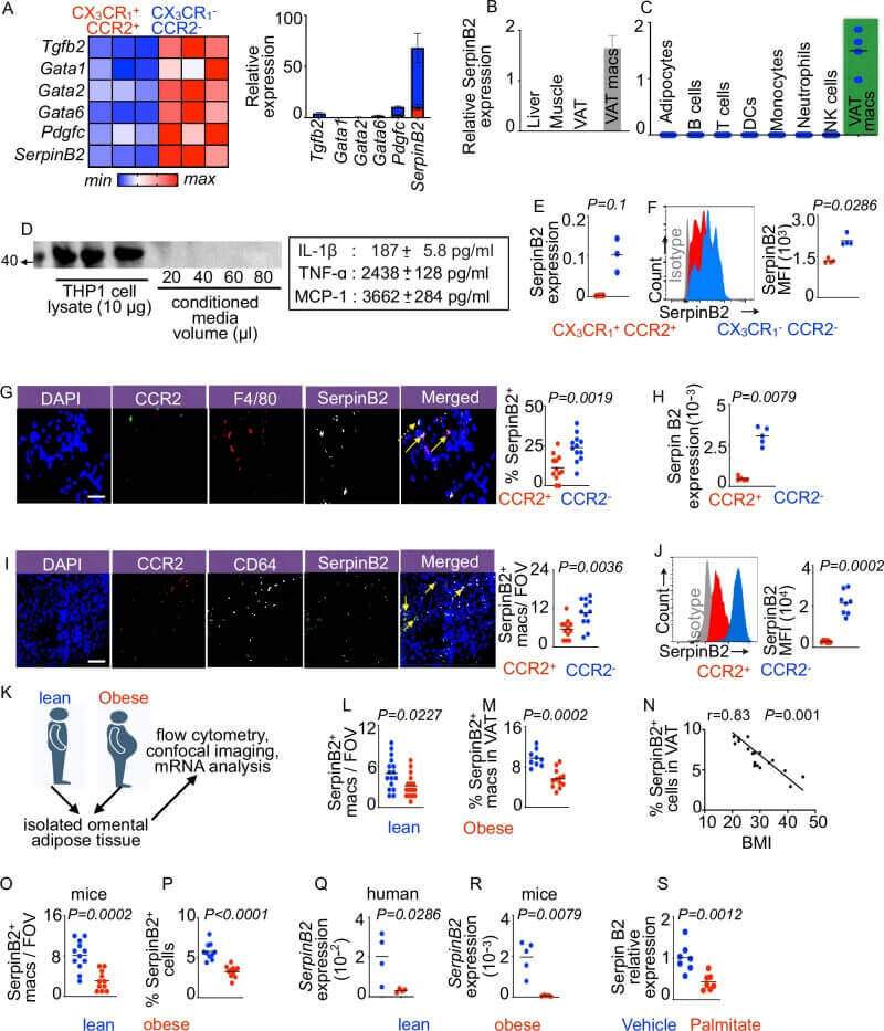 Fig.3 SerpinB2 is highly expressed in resident adipose macrophages and declines with obesity related inflammation. (Vasamsetti, et al., 2026)
Elevated interferon-gamma (IFN-γ) in obese adipose tissue drives SerpinB2 silencing. Mechanistically, IFN-γ induces the transcriptional suppressor Ikaros to bind the SerpinB2 promoter, suppressing its transcription, whereas the Ikaros inhibitor lenalidomide rescues expression. Consequently, myeloid-specific IFN-γ receptor 1 (Ifngr1) deficiency prevents TRM apoptosis and improves systemic metabolism on HFD. Conversely, SerpinB2 deletion causes mitochondrial dysfunction, cytochrome c leakage, and caspase-3-mediated cell death.
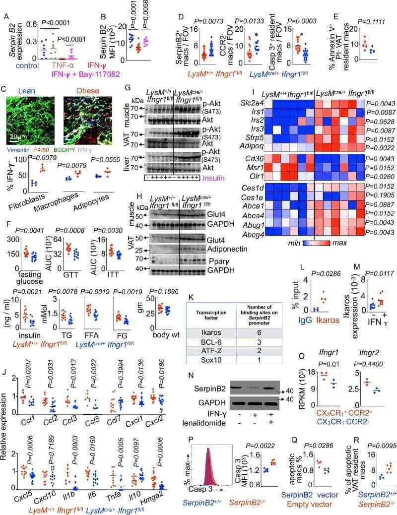 Fig.4 IFN γ signaling reduces SerpinB2 expression and promotes macrophage apoptosis in obese adipose tissue. (Vasamsetti, et al., 2026)
Without SerpinB2, mitochondrial cytochrome c leaks into the cytoplasm. Healthy TRMs are normally enriched with antioxidant genes (e.g., Sod3, Gst family, Mgst1, Mt1/2, Gpx7, Me1, Idh2) and the glutamic acid transporter Slc7a13 to synthesize glutathione (GSH). In obesity, this antioxidant defense declines, correlating with glucose intolerance.
SerpinB2 deficiency reduces these antioxidants, generating mitochondrial reactive oxygen species (ROS) that trigger apoptosis. Scavenging mitochondrial ROS with mitotempol prevents cytochrome c leakage and apoptosis, confirming ROS as the primary driver of cell death.
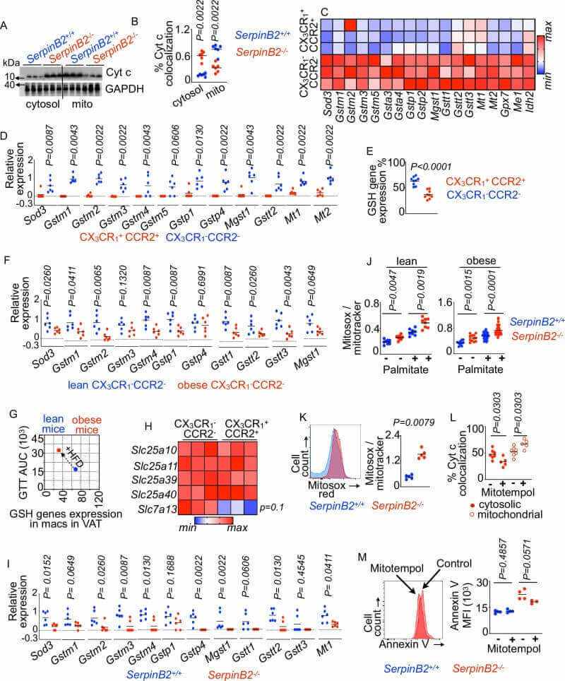 Fig.5 Increased mitochondrial oxidative stress drives apoptosis in SerpinB2-deficient macrophages. (Vasamsetti, et al., 2026)
Obese macrophages exhibit hyperactive oxygen consumption rates (OCR) that fuel inflammatory cytokine release. Inhibiting mitochondrial respiration in myeloid cells (LysM-Cre/Cox10-fl/fl) suppresses this inflammation and improves insulin sensitivity. Conversely, SerpinB2 deficiency accelerates OCR and exacerbates inflammation, whereas its overexpression suppresses inflammation.
Myeloid-specific SerpinB2 deficiency (LysM-Cre/SerpinB2-fl/fl) triggers systemic insulin resistance, glucose intolerance, and elevated lipids. Hyperinsulinemic-euglycemic clamps showed a reduced glucose infusion rate (GIR) and impaired suppression of hepatic glucose production, alongside compromised tissue insulin signaling. Remarkably, administering IL-4 (naturally secreted by TRMs) completely reversed this metabolic dysfunction.
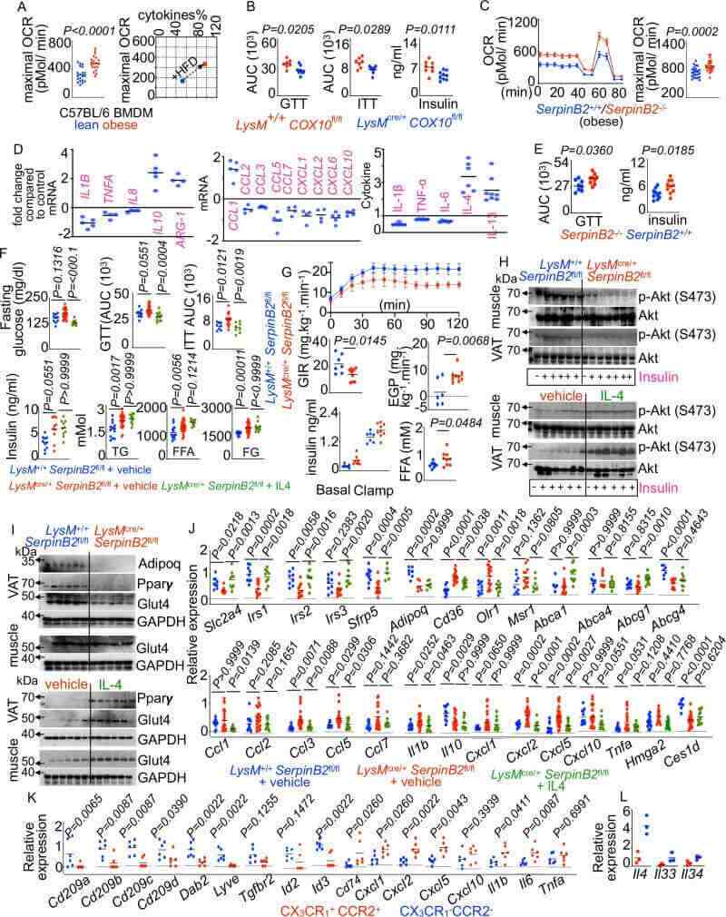 Fig.6 SerpinB2 deficiency enhances inflammatory activity and impairs metabolic regulation in obesity. (Vasamsetti, et al., 2026)
TRM depletion or myeloid SerpinB2 deficiency caused VAT expansion, adipocyte hypertrophy, and altered cytoskeletal remodeling. Adipocytes adjacent to CCR2- TRMs remained smaller, as TRMs express low levels of the hypertrophic factor Il1b and high levels of the inhibitor Tgfb2.
These size-limiting effects are mediated by GSH; the GSH precursor NAC blocked 3T3-L1 adipocyte differentiation in vitro. In vivo, oral NAC supplementation in obese LysM-Cre/SerpinB2-fl/fl mice reduced body weight, restored glucose tolerance, and rescued the TRM population from apoptosis. Conditioned media experiments confirmed that GSH-stimulated TRM secretions directly prevent pathogenic adipocyte hypertrophy.
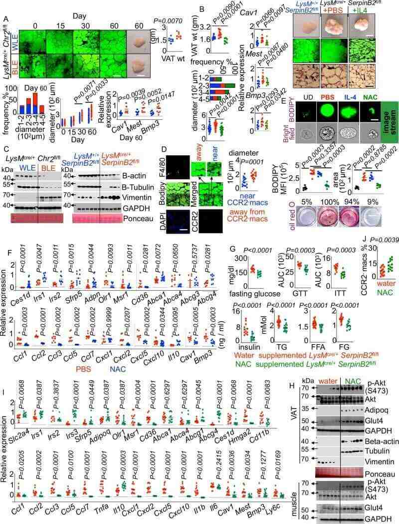 Fig.7 Glutathione signaling limits adipocyte enlargement and visceral fat expansion during obesity. (Vasamsetti, et al., 2026)
This study reveals a key mechanism linking chronic inflammation to metabolic dysfunction, showing how the loss of protective TRMs drives systemic insulin resistance. By identifying the IFN-γ-Ikaros-SerpinB2-GSH axis, the authors highlight new therapeutic targets for type 2 diabetes and obesity-associated metabolic syndrome.
Therapies designed to inhibit the transcriptional suppressor Ikaros, block IFN-γ signaling in myeloid cells, or support mitochondrial antioxidant pathways could help preserve protective TRMs during chronic inflammation.
Furthermore, the discovery that oral administration of the GSH precursor NAC restores the resident macrophage pool and improves insulin sensitivity offers a practical, translatable therapeutic strategy. This mitochondrial rescue approach could also be applied to other inflammatory diseases where tissue-resident macrophages are lost, such as myocardial infarction and Alzheimer's disease.
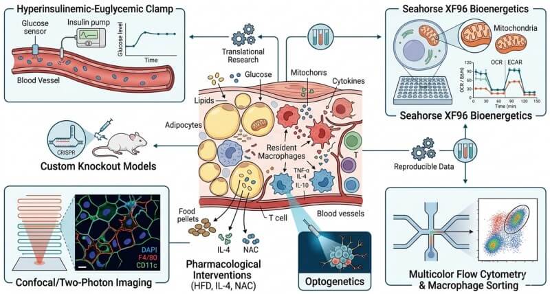
To accelerate your immunometabolism research, Protheragen offers gold-standard preclinical services modeled after this study's key methodologies. We provide hyperinsulinemic-euglycemic clamps to assess insulin sensitivity, Seahorse XF96 bioenergetics for real-time mitochondrial analysis, and multicolor flow cytometry for resident macrophage sorting. Additionally, we support advanced confocal/two-photon tissue imaging, custom knockout models, optogenetics, and pharmacological interventions (HFD, IL-4, or NAC) to deliver reproducible translational data.
Reference
June 22, 2026
June 22, 2026
November 7, 2025
Copyright © Protheragen. All rights reserved.