Guardians of Metabolic Balance: How SerpinB2 Shields Tissue-Resident Macrophages from Mitochondrial Collapse in Chronic Inflammation
June 22, 2026

June 22, 2026
Obesity (OB) is characterized by the excessive accumulation of adipose tissue due to increased nutrient intake and insufficient energy expenditure. It represents a massive global health crisis, with high body mass index (BMI) values causing approximately 5 million non-communicable disease deaths annually. A key consequence of OB is insulin resistance (IR), a metabolic state where insulin-targeting tissues exhibit reduced responsiveness despite high physiological levels of insulin. To address the molecular gaps connecting fat accumulation to impaired insulin action, a comprehensive narrative review was published in the journal Molecular Biology Reports (2026) by a team of specialist researchers. While historical literature often addresses individual treatment modalities or isolated pathways, this specific paper bridges these gaps by providing an integrated synthesis of molecular mechanisms, adipose tissue dysfunction, chronic inflammation, oxidative stress, and multimodal therapeutic strategies.
Under healthy physiological conditions, insulin regulates glucose metabolism by binding to the insulin receptor, triggering tyrosine phosphorylation of insulin receptor substrate 1 (IRS-1). This recruits phosphatidylinositol 3-kinase (PI3K) to convert phosphatidylinositol 4,5-bisphosphate (PIP2) into phosphatidylinositol 3,4,5-trisphosphate (PIP3). Consequently, phosphoinositide-dependent kinase 1 (PDK1) and mechanistic target of rapamycin complex 2 (mTORC2) phosphorylate protein kinase B (Akt). Activated Akt drives glucose transporter 4 (GLUT4) translocation for glucose uptake and inactivates glycogen synthase kinase-3 (GSK-3) to promote glycogen synthase (GS) activity.
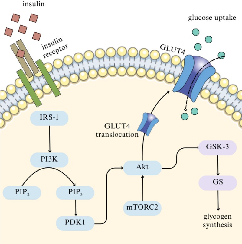 Fig.1 Insulin signalling regulates glucose uptake and glycogen production through coordinated activation of IRS1, PI3K, Akt, and GLUT4 pathways. (Choo, et al., 2026)
In OB, this highly regulated system breaks down through several key pathophysiological mechanisms:
Adipocyte hypertrophy exceeds tissue buffering capacity, triggering excess lipolysis and releasing free fatty acids (FFAs) that cause lipotoxicity in the liver, pancreas, and skeletal muscle. Enlarged cells also suffer from adipocyte hypoxia, which stabilizes hypoxia-inducible factor-1α (HIF-1α). This promotes the polarization of macrophages from the anti-inflammatory M2 phenotype to the pro-inflammatory M1 phenotype via Toll-like receptor 4 (TLR4). M1 macrophages secrete pro-inflammatory cytokines like tumour necrosis factor α (TNF-α) and interleukin-6 (IL-6), while reducing insulin-sensitizing adipokines like adiponectin. Hypertrophy also upregulates exosomal miR-802-5p, silencing heat shock protein 60 (HSP60) and inducing oxidative stress. This oxidative stress is driven by mitochondrial dysfunction and nicotinamide adenine dinucleotide phosphate (NADPH) oxidase (NOX) enzymes like NOX4, generating excessive reactive oxygen species (ROS) from the electron transport chain (ETC).
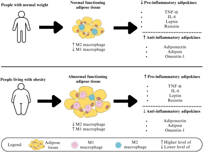 Fig.2 Obesity alters adipokine balance by increasing inflammatory macrophages and reducing anti-inflammatory mediators in adipose tissue. (Choo, et al., 2026)
As shown in Fig. 3, the nuclear factor-kappa B (NF-κB) pathway is activated by hypoxia, ROS, endoplasmic reticulum (ER) stress (via the Unfolded Protein Response (UPR) involving inositol-requiring enzyme 1 (IRE1), protein kinase RNA-like ER kinase (PERK), and activating transcription factor 6 (ATF6)), FFAs, and cytokines. These stimuli activate the IkappaB Kinase (IKK) complex. Once active, NF-κB drives the transcription of TNF-alpha and IL-6, which activate serine kinases like inhibitory kappa B kinase beta (IKKβ) and c-Jun N-terminal kinase (JNK). These kinases phosphorylate IRS-1 on serine residues (e.g., Ser307), blocking critical tyrosine phosphorylation. Concurrently, Suppressors of cytokine signalling (SOCS) proteins, such as SOCS1 and SOCS3, bind to IRS-1, inactivating downstream PI3K/Akt signalling, halting GLUT4 translocation, and impairing glycogen synthesis.
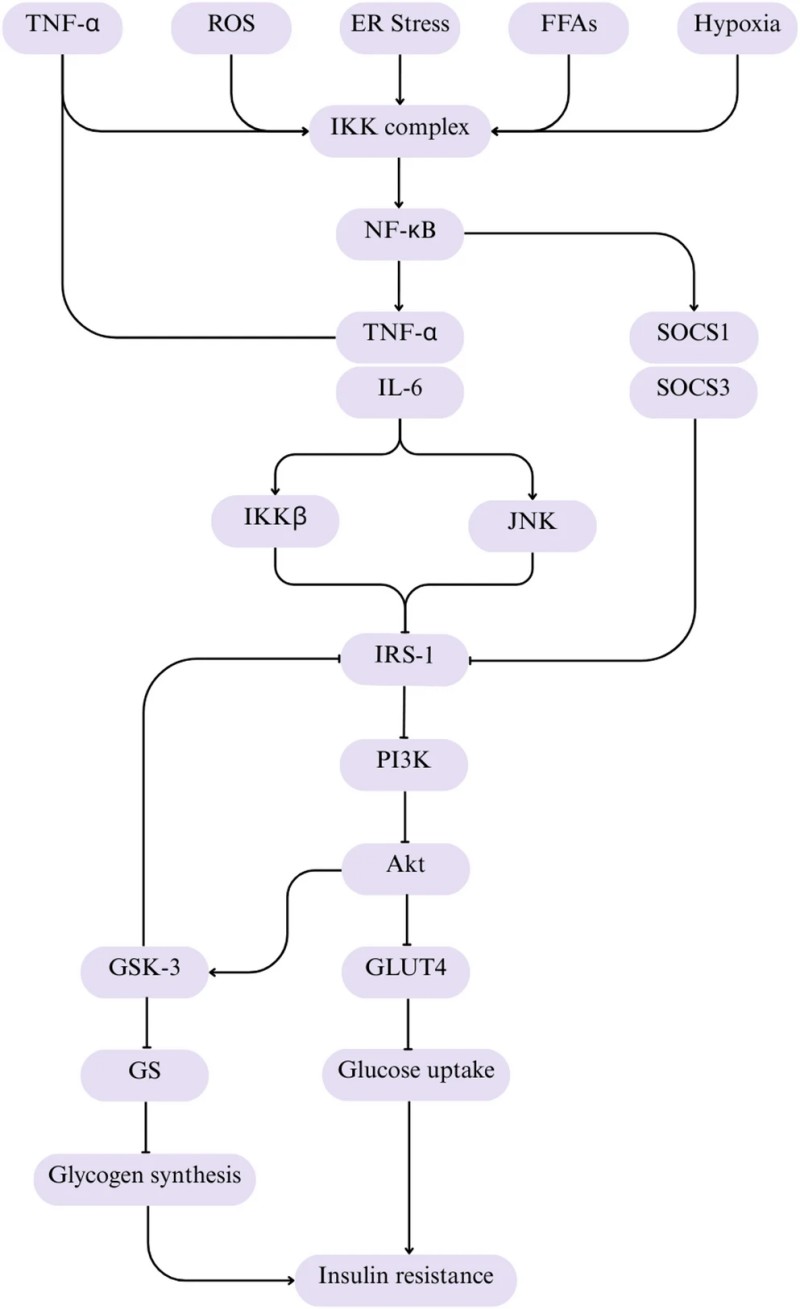 Fig.3 Inflammatory activation of NFκB disrupts insulin signalling and promotes the development of insulin resistance. (Choo, et al., 2026)
To counter these dysregulations, the review outlines three therapeutic pillars:
Furthermore, cutting-edge therapies offer promising precision management:
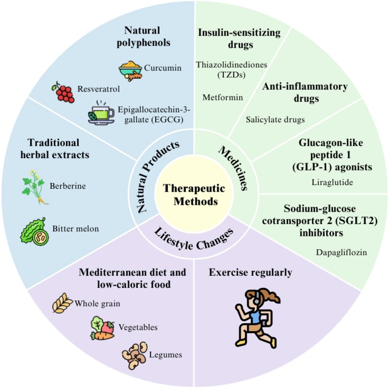 Fig.4 Multiple therapeutic strategies improve obesity related insulin resistance by targeting inflammation, metabolism, and insulin sensitivity. (Choo, et al., 2026)
The translational significance of this research lies in its transition from a generalized treatment model to a highly personalized and integrated medical strategy for addressing metabolic syndrome. By mapping the exact molecular checkpoints where inflammation, adipocyte hypoxia, and oxidative stress cross paths with the insulin signaling cascade, this review provides a roadmap for developing targeted, synergistic therapies. Furthermore, the paper highlights how therapeutic outcomes can be maximized by incorporating pharmacogenomics to analyze genetic variations, such as identifying individuals with specific PRPF31, CPA6, or STAT3 genes who respond well to metformin, versus those with SLC22A2 gene variants who require alternative treatments. When combined with advanced digital health technologies, AI predictive models, and gene-editing platforms targeting markers like NRIP1 and FABP4, these scientific insights can be successfully translated into scalable, real-world clinical interventions that improve long-term metabolic recovery worldwide.
For researchers and clinical organizations aiming to explore novel therapeutic targets, metabolic profiling, or preclinical models of metabolic disease, Protheragen offers comprehensive analytical services, custom in vivo disease modeling, and advanced cellular assay services to accelerate your drug discovery and translation pipeline.
Reference
June 22, 2026
June 22, 2026
November 7, 2025
Copyright © Protheragen. All rights reserved.