
Macrophage Cell Isolation and Culture Service
InquiryObesity is no longer viewed simply as an energy storage problem. It is a chronic, low-grade inflammatory state driven largely by the immune cells infiltrating expanding fat pads. Within adipose tissue, macrophages change dramatically during weight gain. They shift from a protective, anti-inflammatory phenotype to a pro-inflammatory state that secretes massive amounts of cytokines like TNF-ɑ and IL-6. This driving force behind insulin resistance and metabolic syndrome makes macrophages prime targets for novel cell therapies.
Macrophage Cell Isolation and Culture Service in Macrophage Cell Therapy Development
Developing effective preclinical cell therapies requires high-quality, highly functional macrophages isolated directly from complex tissue environments. Protheragen offers specialized services designed specifically to isolate and culture these delicate cells from adipose tissues and other obesity-affected organs. We handle the complex tissue digestion and sorting processes, so your team can focus entirely on downstream therapeutic testing.
Core Technologies
Isolating macrophages from lipid-dense adipose tissue presents unique physical and biological challenges. Standard isolation methods often damage cells or fail to separate target cells from floating adipocytes. Protheragen has optimized a suite of proprietary platforms to yield viable, unactivated cells from metabolic tissue matrices.
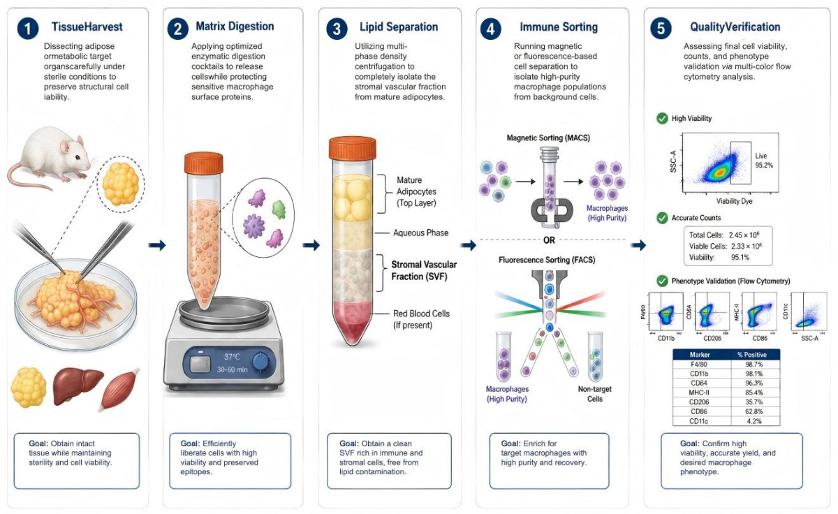
Optimized collagenase and dispase cocktails break down the extracellular matrix without cleaving vital cell surface receptors.
Positive or negative selection using verified antibodies targeting CD11b, F4/80, and CD11c yields pure macrophage fractions.
Specialized media layers remove excess lipid droplets and free-floating mature adipocytes that typically contaminate downstream assays.
Serum-free and low-endotoxin culture conditions preserve the baseline activation state, preventing unwanted ambient polarization before your experiments begin.
Solution Scope
Our capabilities span the entire front-end pipeline of preclinical macrophage assay development. We customize each project to match the precise metabolic model and strain your study relies upon.
- Primary Source Extraction & Isolation
We regularly isolate macrophages from a wide variety of tissue microenvironments affected by chronic metabolic disease. This includes visceral adipose tissue (VAT), subcutaneous adipose tissue (SAT), liver tissues (Kupffer cell populations), and skeletal muscle. We work with standard preclinical rodent models, including diet-induced obesity (DIO) mice, ob/ob, and db/db models.
- Phenotypic Characterization and Profiling
Understanding your starting population is essential. We provide comprehensive baseline characterization using multi-marker flow cytometry pan-macrophage markers alongside detailed polarization panels. This includes assessing the balance between pro-inflammatory and anti-inflammatory markers to accurately map the inflammatory state of your sample tissue.
- In Vitro Culturing and Polarization Assays
We maintain primary cells in specialized, ultra-low endotoxin conditions to prevent spontaneous activation. Our team can direct cells toward specific states using precise combinations of cytokines or metabolic triggers like free fatty acids. This lets you test how your therapeutic candidates alter macrophage behavior in a highly controlled environment.
- Functional Testing and Preclinical Validation
We offer functional assays to evaluate phagocytic capacity, chemotaxis response, and detailed cytokine secretion profiles. These metrics help you confirm whether your target therapeutics can successfully modulate macrophage activity, reduce tissue inflammation, or improve metabolic function in vivo.
Inquire with our technical team to design an isolation protocol.
Workflow
Our streamlined, high-efficiency extraction protocol is built to protect cell health while systematically isolating target populations from dense metabolic tissues.
Fields of Application
By delivering highly pure, unactivated macrophage populations, our platform serves as a critical foundational layer across several distinct areas of translational metabolic research.
- Preclinical Drug Screening
Screen small molecule candidates, biologics, or RNA therapies designed to block pro-inflammatory signaling pathways or accelerate the resolution of chronic tissue inflammation. - Metabolic Pathway Discovery
Map out how lipid accumulation, hypoxia, and changes in the local extracellular matrix alter macrophage behavior, helping you identify novel therapeutic targets. - Adoptive Cell Therapy Modeling
Develop and optimize engineered macrophage candidates designed to clear tissue debris, resolve fibrosis, or restore metabolic balance when re-introduced into Preclinical Models.
Advantages
Partnering with Protheragen means bypassing the steep learning curve and high failure rates typically associated with processing delicate, lipid-heavy immune samples.
Exceptional Yields from Challenging Tissues
Adipose tissue is notorious for low immune cell yields and high lipid interference. Protheragen leverages proprietary density-gradient techniques that consistently deliver two to three times more viable macrophages per gram of fat tissue compared to standard generic tissue kits. This maximizes your data output from expensive animal cohorts.
Preserved Baseline Polarization States
Standard isolation techniques frequently trigger accidental activation due to rough mechanical handling or high endotoxin levels. Our specialized, low-stress workflows protect cell integrity, ensuring the macrophages you receive accurately reflect the true inflammatory profile of the donor tissue rather than artifacts from the lab process.
Extensive Preclinical Experience
Our scientific team has completed numerous isolation projects across a wide variety of metabolic disease models. In past evaluation work, our isolated cells demonstrated excellent functional responses in downstream co-culture models, as backed by our internal data tracking viability over extended assay windows.
Connect with our specialists to receive a transparent project outline.
Publication Data
Title: Obesity-induced senescent macrophages activate a fibrotic transcriptional program in adipocyte progenitors
Journal: Life Science Alliance, 2022
DOI: http://doi.org/10.26508/lsa.202101286
Summary: Single-cell RNA-seq on mouse adipose tissue reveals high-fat diet triggers senescent CD9+ macrophages with blunted phagocytosis. These cells secrete osteopontin, driving PDGFRα/β double-positive adipocyte progenitors to boost ECM production, block adipogenesis and fuel obesity-linked white adipose tissue fibrosis.
Key Findings
- Primary ECM-producing progenitor cell identified
Single-cell RNA sequencing of mouse epididymal white adipose tissue (eWAT) confirms MSC1 mesenchymal stromal cells co-expressing PDGFRα and PDGFRβ are the main extracellular matrix (ECM) producers. A high-fat diet (HFD) expands this progenitor pool and upregulates collagen fibrotic genes; PDGFRβ downregulation pushes progenitors toward mature adipocyte differentiation. - Obesity generates dysfunctional senescent CD9+ macrophages
HFD drives enrichment of CD9+ macrophages within adipose crown-like structures. These immune cells display canonical senescence markers (p16, p21, SA-β-gal), lose normal phagocytic capacity, and secrete a pro-fibrotic, pro-inflammatory secretory phenotype. - Osteopontin (OPN/Spp1) acts as core intercellular fibrotic signal
NicheNet ligand-receptor analysis singles out osteopontin, exclusively overexpressed by senescent CD9+ macrophages under HFD, as the key mediator communicating with PDGFRα/β progenitors. - OPN + PDGF-BB synergize to lock progenitors in a fibrotic state
In vitro testing shows combined OPN and PDGF-BB treatment amplifies progenitor cell proliferation, elevates fibrotic gene expression, and fully suppresses adipogenesis. This traps immature ECM-making progenitors in obese fat, worsening adipose tissue scarring. - Translational therapeutic insight
The study establishes obesity-induced macrophage senescence as a root driver of adipose fibrosis, highlighting OPN and CD9+ macrophages as actionable targets for treating obesity-associated metabolic and inflammatory diseases.
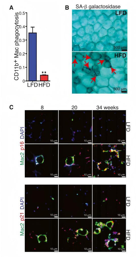 Fig.1 How senescent CD9+ macrophages trigger adipose tissue fibrosis in obesity. (Rabhi, et al., 2022)
Customer Review
Overcoming Lipid Contamination Obstacles in High-Throughput Downstream Assays
"Isolating clean macrophage fractions from our long-term DIO mouse fat pads was a major bottleneck for our team. The lipid contamination was ruining our downstream PCR work. Protheragen took over the extraction process and delivered incredibly clean, high-purity cell fractions. This saved us months of trial-and-error, and we are already planning our next batch of tissue runs with them."
Dr. A. V., Scientific Researcher
Accelerating Target Validation via Standardized Polarization Frameworks
"We needed a reliable partner to test our engineered cell therapy candidates in a primary tissue setting. The team at Protheragen did an excellent job setting up the polarization assays and provided clear flow cytometry data that matched our expectations perfectly. Their technical communication is outstanding, and we look forward to continuing this collaboration as our program advances."
Dr. D. I., Lab Manager
Frequently Asked Questions
-
Can you isolate macrophages from heavily fibrotic adipose tissue samples?
Yes. We adjust our digestion protocols with specific collagenase blends to effectively break down the dense extracellular matrix found in long-term diet-induced obesity models without losing cell surface markers.
-
What typical purities do you achieve for adipose tissue macrophages?
Our magnetic sorting protocols consistently deliver purities greater than 90% for standard macrophage populations, which we verify via flow cytometry before shipment or downstream assays.
-
How do you prevent macrophages from altering their polarization during extraction?
We keep all steps cold, work quickly, and use endotoxin-free reagents and specialized media that lack activating factors to maintain the cell's native state.
-
Are you able to process tissues from custom or unique genetic mouse models?
Absolutely. You can ship tissues directly to our facility using our optimized cold-transport protocols, or we can handle the model housing and tissue harvesting in-house.
-
What functional assays can you run after isolating the cells?
We regularly perform cytokine profiling, phagocytosis tracking, lipid accumulation assays, and metabolic flux analysis using specialized extracellular analyzers.
-
Can we get both M1-like and M2-like subsets isolated separately from the same tissue?
Yes, we can perform sequential magnetic sorting or multi-channel fluorescence-activated sorting to isolate distinct sub-populations based on specific marker combinations.
-
How long can these primary tissue macrophages be maintained in culture?
Primary tissue-resident macrophages do not proliferate extensively. We optimize media formulations to maintain high viability and functional responses for up to 7–10 days depending on the assay design.
-
Does your service include isolation from non-adipose metabolic tissues?
Yes, we frequently isolate tissue macrophages from liver, skeletal muscle, and spleen samples from metabolic and obese mouse models.
-
How do you ensure there is no adipocyte contamination in our cell cultures?
We use multi-stage density washes alongside strict magnetic negative selection to ensure lipid droplets and adipocyte debris are thoroughly removed from the final cell suspension.
Contact Us
Targeting chronic tissue inflammation by modulating macrophage behavior holds incredible promise for the future of metabolic medicine. Succeeding with these complex programs requires reliable, reproducible cell isolation techniques that preserve natural cellular behavior. Protheragen provides the specialized technology, validated workflows, and deep scientific expertise needed to deliver high-quality primary cell preparations from challenging metabolic environments. Let us manage the complexities of tissue processing so your team can focus on developing breakthrough therapeutics. Welcome to Contact Protheragen today to discover how our high-performance molecular tools can accelerate your discovery process.
Reference
- Rabhi, N.; et al. Obesity-induced senescent macrophages activate a fibrotic transcriptional program in adipocyte progenitors. Life Science Alliance. 2022, 5(5), e202101286 (CC BY 4.0).
All of our services and products are intended for preclinical research use only and cannot be used to diagnose, treat or manage patients.