
Glucose Metabolic Dynamics and Rate Analysis
InquiryGlucose metabolism is the cornerstone of cellular bioenergetics and systemic homeostasis. In preclinical research, relying on static "snapshot" measurements—such as blood glucose levels or mRNA expression—often fails to capture the true functional state of an organism.
High-Resolution Glucose Kinetics & Flux Analysis for Preclinical Research
Protheragen provides a sophisticated suite of glucose metabolic dynamics and rate analysis services designed to move beyond static data. By measuring the actual flux and kinetic rates of glucose appearance (Ra) and disappearance (Rd), we empower researchers to identify the precise mechanisms of metabolic dysregulation. Our services focus exclusively on preclinical models, offering high-resolution insights into glycolysis, oxidative phosphorylation, and whole-body glucose turnover.
Core Technologies
To deliver industry-leading accuracy, Protheragen utilizes a multi-platform approach that integrates real-time cellular monitoring with systemic tracer kinetics:
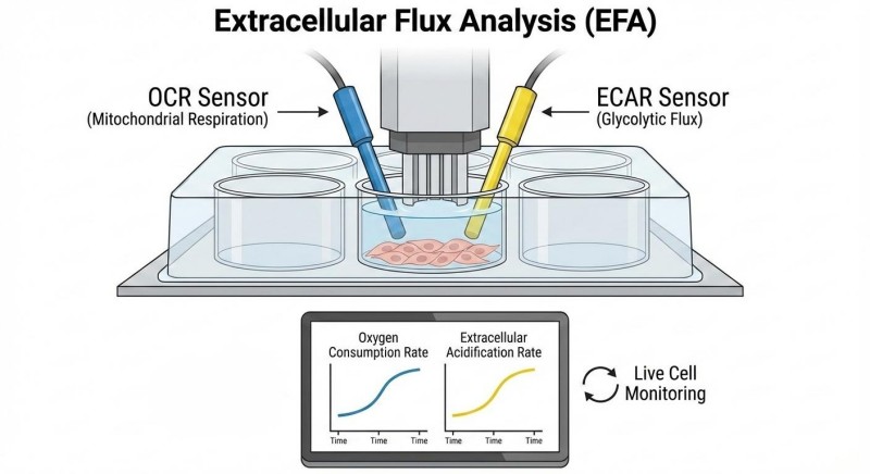
- Extracellular Flux Analysis (EFA)
Utilizing state-of-the-art sensor technology to simultaneously measure the oxygen consumption rate (OCR) and extracellular acidification rate (ECAR) in live cells or ex vivo tissue slices. This provides a direct readout of mitochondrial respiration and glycolytic flux.
- Stable Isotope-Resolved Metabolomics (SIRM)
We employ [U-13C]-glucose and other non-radioactive tracers. By using liquid chromatography-tandem mass spectrometry (LC-MS/MS) and gas chromatography-mass spectrometry (GC-MS), we track the incorporation of labeled carbons into downstream metabolites (e.g., pyruvate, lactate, TCA cycle intermediates), allowing for precise metabolic pathway reconstruction.
- Hyperinsulinemic-Euglycemic Clamp
The gold standard for assessing insulin sensitivity in vivo. This technology allows for the independent quantification of hepatic glucose production and peripheral glucose uptake under controlled steady-state conditions.
- Kinetic Modeling and Simulation
Advanced mathematical models (such as the minimal model) are used to derive the insulin sensitivity index (SI) and glucose effectiveness (SG) from dynamic glucose tolerance tests.
Service Scope
Protheragen offers a broad spectrum of preclinical glucose analysis services tailored to various therapeutic areas:
- Whole-Body Glucose Turnover
Measuring Ra and Rd using stable isotope tracers to determine systemic metabolic health.
- In Vivo Tissue-Specific Uptake
Quantifying glucose disposal in skeletal muscle, adipose tissue, and the brain.
- Cellular Glycolytic Rate Assays
Real-time quantification of basal glycolysis, compensatory glycolysis, and glycolytic capacity in live cell populations.
- Substrate Oxidation Profiles
Investigating the "fuel flexibility" of cells—determining how they shift between glucose, fatty acids, and glutamine under metabolic stress.
- Hepatic Glucose Production (HGP)
Assessing the liver's contribution to hyperglycemia in diabetic and obese models.
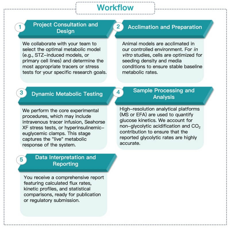
Workflow
Our streamlined service workflow ensures that every preclinical project receives rigorous scientific oversight from design to data delivery:
Fields of Application
The glucose metabolic dynamics and rate analysis provided by Protheragen serve as a critical diagnostic and evaluative tool across a diverse range of preclinical therapeutic areas, enabling researchers to decode the functional metabolic shifts that underpin complex disease states.
- Diabetes and Obesity Research: Identifying the primary drivers of insulin resistance and evaluating the efficacy of glucose-sensitizing agents.
- Immunometabolism: Analyzing how metabolic reprogramming drives T-cell activation and immune response dynamics.
- Cardiovascular Disease: Evaluating glucose utilization in failing hearts and assessing the metabolic impact of ischemia/reperfusion injury.
- Toxicology: Screening drug candidates for off-target metabolic effects or mitochondrial toxicity early in the development pipeline.
Advantages
Choosing Protheragen means accessing a level of precision that standard labs cannot match. Our glucose metabolic dynamics and rate analysis service is distinguished by:
- Kinetic Accuracy
We go beyond static concentrations to measure true metabolic turnover. Our "flux" analysis distinguishes whether metabolite changes are driven by increased production or decreased consumption, providing a clear map of drug impact.
- High Sensitivity
Get high-resolution data with 3x fewer cells. Our optimized extracellular flux systems are purpose-built for precious primary cells and organotypic slices, maximizing data from your most limited samples.
- Validated Preclinical Models
We utilize "published data" standards to ensure our models (from high-fat diet rodent models to complex 3D organoids) translate effectively to biological reality.
- Label-Free Monitoring
Our real-time assays provide dynamic kinetic data without the need for radioactive labels or destructive sampling, preserving the biological integrity of your samples throughout the experiment.
Contact Our Team for More Information and to Discuss Your Project.
Publication Data
Title: Live-cell metabolic analyzer protocol for measuring glucose and lactate metabolic changes in human cells
Journal: STAR Protocols., 2025
DOI: https://doi.org/10.1016/j.xpro.2024.103518
Summary: Understanding glycolysis-related metabolic conditions is critical for advancing cancer and regenerative medicine treatments. This paper details a protocol for using the LiCellMo live-cell metabolic analyzer to continuously measure glucose consumption and lactate production in cultured human cells—addressing the limitation of traditional extracellular flux analysis, which only captures metabolic snapshots. LiCellMo leverages enzyme-based biosensors to enable real-time, non-destructive detection of extracellular glucose and lactate, with high sensitivity and specificity. The protocol covers cell seeding (for SH-SY5Y and other common cell types), calibration with two specialized buffers, inhibitor administration (e.g., PFK, GLS1, CPT1 inhibitors), and data analysis to calculate metabolic rates. Validated in experiments, the tool provides reliable insights into metabolic pathway activity, supporting metabolic research, drug development, and biomedical studies across cancer biology, neurobiology, and reproductive medicine.
Key Findings
- Tool Advantage: LiCellMo enables real-time, non-destructive, high-sensitivity measurement of glucose/lactate metabolism in human cells, surpassing traditional snapshot methods.
- Detection Mechanism: Relies on enzyme electrodes (glucose/lactate dehydrogenase) to convert metabolic changes into quantifiable electric signals.
- Inhibitor Test Results:
- PFK inhibitors (glycolysis): Reduce glucose consumption and lactate production without cell death.
- GLS1 inhibitors (glutaminolysis): Decrease lactate production with minimal glucose consumption change.
- CPT1 inhibitors (fatty acid metabolism): Increase lactate production while glucose consumption remains stable.
- Protocol Key Points: Specifies cell seeding densities (e.g., 50k SH-Sy5Y cells/well), two calibration buffers (6mM/12mM lactate), 1-minute cycle data recording, and normalization to cell count/protein.
- Applications: Supports metabolic research, drug screening, and cancer/regenerative medicine studies.
- Troubleshooting: Provides solutions for sensor drift, data recording failures, software errors, etc.
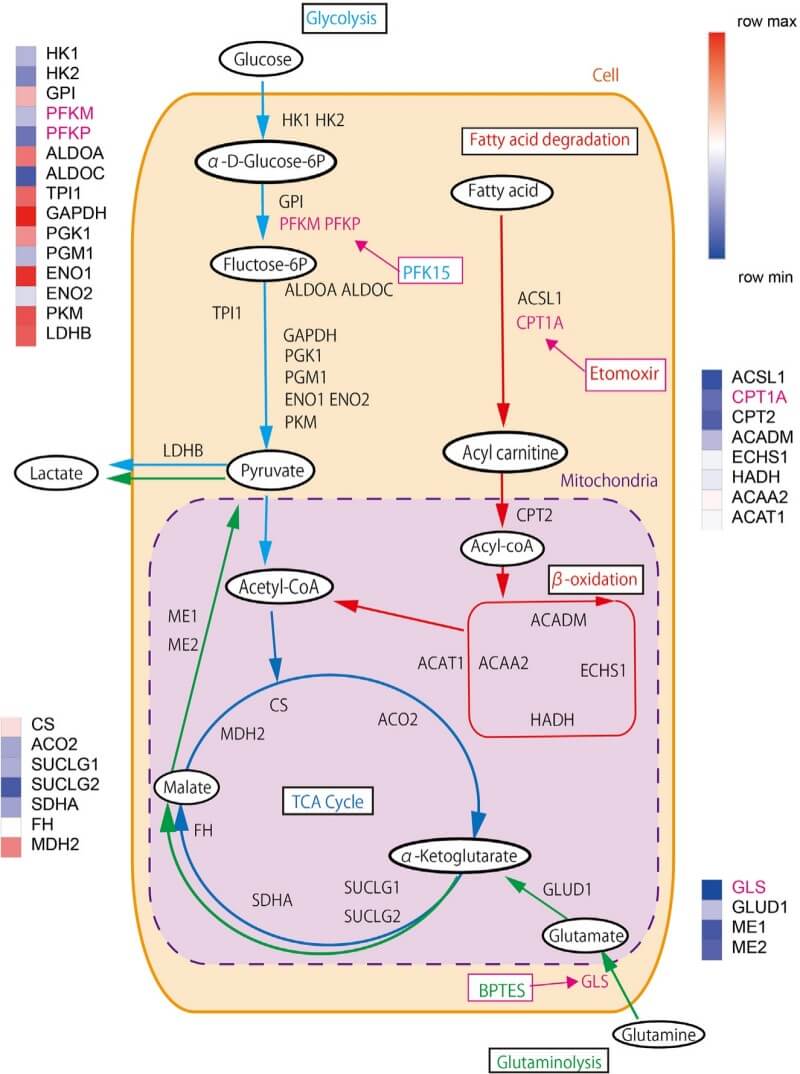 Fig.1 iMPAQT analysis of metabolic enzyme quantities in SH-Sy5Y cells—linking glycolysis, glutaminolysis, and fatty acid metabolism pathways to targeted inhibitors. (Kenji, et al., 2025)
Customer Review
Case Study: Mechanistic Validation of Insulin Sensitizers in Peripheral Tissues
"The team at Protheragen transformed our understanding of our lead compound. We initially thought the drug was affecting insulin secretion, but their hyperinsulinemic-euglycemic clamp and tracer analysis proved the mechanism was actually centered on peripheral glucose disposal. Their expertise in preclinical metabolic flux is unparalleled."
Dr. J. Y., Biotech Lead Generation Group
Technical Spotlight: Precision Bioenergetics in Primary Cell Metabolic Profiling
"Protheragen provided the precise kinetic data we needed for our latest publication. Their ability to run high-sensitivity Seahorse assays on our limited primary cell samples was a game-changer. We are already planning our next series of in vivo studies with their metabolic team."
r. Z. D., Department of Metabolic Research
Frequently Asked Questions
-
Why should I measure glucose flux instead of just checking blood sugar?
Blood sugar is a static pool. A normal blood sugar level could hide a state where both production and uptake are pathologically high. Flux analysis reveals the underlying dynamics that a single blood test misses.
-
What species can you perform these metabolic studies in?
Protheragen focuses on rodent models (mice, rats, etc) and in vitro systems. Please contact us for a full list of supported preclinical species.
-
What is the benefit of using stable isotopes over radioactive tracers?
Stable isotopes like 13C are non-radioactive, safer for laboratory environments, and allow for "labeling depth"—tracing the carbon atoms through multiple metabolic pathways simultaneously via mass spectrometry.
-
How do you handle non-glycolytic acidification in your EFA assays?
We use advanced protocols that account for CO2-derived protons from the TCA cycle, ensuring that the extracellular acidification rate (ECAR) truly reflects glycolytic flux.
-
Is your service compatible with 3D cell models or organoids?
Yes, we have specialized protocols and kits optimized for 3D samples, ensuring oxygen and nutrient gradients are accounted for during metabolic analysis.
-
What is the typical turnaround time for a whole-body glucose turnover study?
While it depends on the complexity, most projects from design to final report are completed within 4–6 weeks.
-
Can I provide my own proprietary compounds for testing?
Absolutely. We frequently perform "acute" or "chronic" dosing studies to see how your candidates affect metabolic dynamics.
-
Do you offer help with the experimental design for NIH or grant-funded projects?
Yes, our specialists have over 20 years of experience and can assist in designing robust experiments that meet the highest peer-review standards.
-
Are the results delivered in a format ready for publication?
Yes, we provide high-resolution figures, detailed methodology sections, and raw data files.
Contact Us
Protheragen provides world-class glucose metabolic dynamics and rate analysis for preclinical research. By integrating stable isotope tracing, extracellular flux analysis, and advanced in vivo clamping techniques, we offer a holistic view of glucose homeostasis. Our data-driven approach helps researchers identify therapeutic targets, validate drug efficacy, and uncover the complex mechanisms of metabolic disease with precision and reliability.
Contact Protheragen for More Information and to Discuss Your Project
Reference
- Kenji M.; et al. Live-cell metabolic analyzer protocol for measuring glucose and lactate metabolic changes in human cells. STAR Protocols. 2025, 6(1):103518. (CC BY 4.0)
All of our services and products are intended for preclinical research use only and cannot be used to diagnose, treat or manage patients.