New Discovery on the Culprit Behind Obesity: How Fat Cells Ignite Inflammation and Metabolic Crisis Through the B2M Protein?
December 3, 2025
June 22, 2026
Maintaining a stable body temperature is a vital homeostatic process in endothermic animals, regulated by brown adipose tissue (BAT) through adaptive thermogenesis. During cold exposure, BAT thermogenesis requires a highly synchronized expansion of its neurovascular niche, specifically the parallel growth of blood vessels and sympathetic nerves. While previous studies detailed how individual adipocytes activate their thermogenic machinery, the molecular mechanisms coordinating this surrounding microenvironmental remodeling remained unclear. Historically, the local stromal vascular fraction was viewed as a passive reservoir, and past research failed to identify the upstream signals driving these synchronized vascular and neural adjustments. This gap was addressed in a landmark Nature Communications study published in early 2026, titled "SLIT3 fragments orchestrate neurovascular expansion and thermogenesis in brown adipose tissue." The researchers leveraged single-cell RNA sequencing (scRNA-seq) and conditional mouse models to investigate how intercellular communication networks drive this tissue-wide adaptation.
Ultimately, these key findings demonstrate a sophisticated, bifurcated communication network where a single progenitor-derived ligand is enzymatically processed to independently yet harmoniously direct both the vascular and neural architectures of the thermogenic niche.
The researchers utilized scRNA-seq to construct a comprehensive map of intercellular ligand-receptor pairs in the cold-acclimated adipose niche. This systematic screen revealed that Slit3 is predominantly and highly expressed within PDGFRα-expressing ASPCs, while its canonical vascular counterpart, ROBO4, is restricted entirely to endothelial cells. Protein analysis confirmed that environmental cold exposure robustly increases SLIT3 protein levels in the tissue.
To test the functional requirement of this pathway in vivo, the team utilized adeno-associated virus (AAV) vectors carrying short hairpin RNA (shRNA) constructs to induce a localized knockdown of Slit3 within the interscapular BAT of adult mice. When subjected to a cold challenge, mice lacking BAT-derived SLIT3 exhibited severe thermogenic deficits, maintaining lower core body temperatures and reduced BAT temperatures. Histological examination via hematoxylin and eosin (H&E) staining showed a severe morphological whitening of the tissue characterized by massive lipid accumulation. At the molecular level, quantitative Western blot and real-time polymerase chain reaction analyses demonstrated a profound down-regulation of uncoupling protein 1 (UCP1) alongside key mitochondrial electron transport chain (ETC) complex proteins. Metabolic cage assessments confirmed a substantial drop in absolute energy expenditure in the knockdown group.
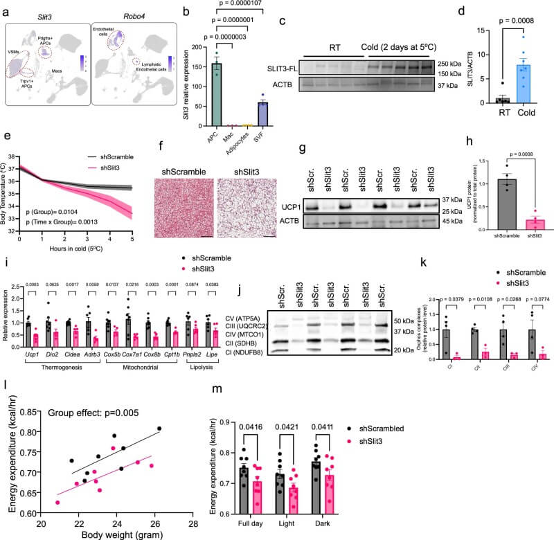 Fig.1 Cold exposure increases Slit3 expression and supports brown fat thermogenic activity. (Serdan, et al., 2026)
Crucially, the investigation proved that these metabolic defects stem from architectural failures within the tissue niche. The depletion of SLIT3 caused a massive reduction in capillary density, which was confirmed by quantifying Isolectin B4 (IB4) positive vascular surface areas. Concurrently, sympathetic neurite density was drastically diminished, as evidenced by a loss of tyrosine hydroxylase (TH) immunofluorescence. Three-Dimensional (3D) tissue clearing via the adipo-clear protocol combined with advanced light-sheet microscopy visually confirmed a near-complete loss of the parenchymal sympathetic nerve network in the absence of the ligand. Strikingly, lineage-specific deletion of Slit3 in PDGFRα-expressing progenitors (using inducible Pdgfra-creERT2;Slit3flox/flox mice) fully recapitulated these thermogenic and neurovascular defects, confirming ASPCs as the indispensable physiological source of SLIT3 in BAT.
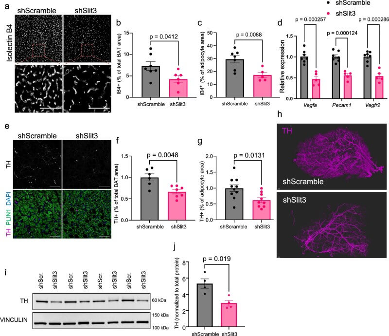 Fig.2 Slit3 deficiency reduces blood vessel growth and sympathetic nerve density in brown fat during cold adaptation. (Serdan, et al., 2026)
Biochemical analyses resolved how this single full-length protein executes two distinct tasks. Using epitope-tagged constructs expressed in progenitor cell lines, the authors proved that full-length SLIT3 is processed into an N-terminal fragment (SLIT3-N) and a C-terminal fragment (SLIT3-C). Mass spectrometry data verified that SLIT3-N remains partially membrane-associated to drive local endothelial proliferation, whereas SLIT3-C diffuses freely into the parenchyma. Pharmacological screening using the small-molecule inhibitor UK383,367 and genetic silencing via small interfering RNA (siRNA) proved that the zinc metalloproteinase BMP1 is the specific enzyme driving this cleavage in vertebrates.
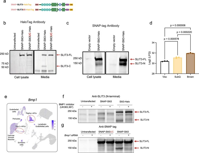 Fig.3 BMP1 cleavage produces active SLIT3 fragments secreted by adipose cells. (Serdan, et al., 2026)
By delivering engineered AAV constructs overexpressing specific fragments in mouse tissue, the authors demonstrated that SLIT3-N selectively expands the IB4-positive capillary network, whereas SLIT3-C specifically stimulates sympathetic innervation and elevates tissue temperature. To identify the receptor mediating the neural effects of SLIT3-C, the team examined candidate receptors. Immunofluorescence and co-immunoprecipitation assays showed that PLXNA1 and ROBO1 localize to TH-positive sympathetic nerves, and that both full-length SLIT3 and SLIT3-C physically interact with the extracellular region of PLXNA1.
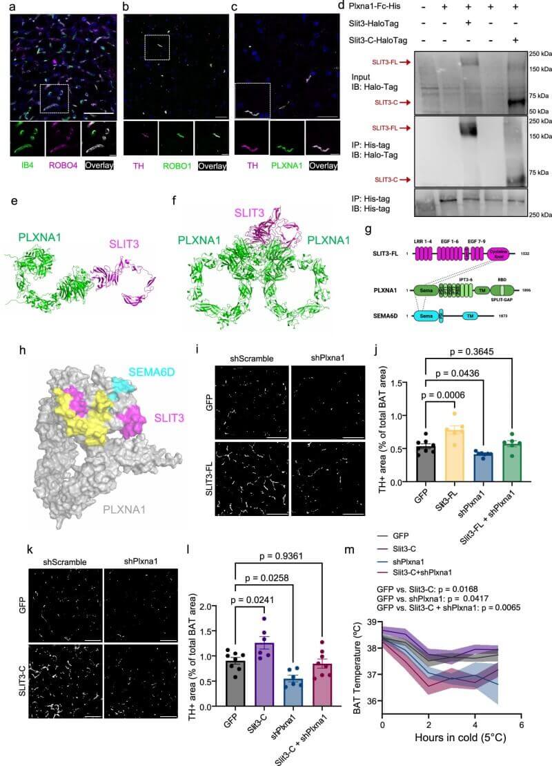 Fig.4 PLXNA1 functions as a receptor for SLIT3 to regulate sympathetic innervation in brown adipose tissue. (Serdan, et al., 2026)
AlphaFold2 structural modeling revealed two distinct interaction modes where the extracellular domain of PLXNA1 binds SLIT3-C as a monomer or dimer, showing a binding footprint that partially overlaps with the SEMA6D interface. In vivo, Plxna1 knockdown in BAT completely blocked the pro-innervation and thermogenic rescue effects of SLIT3-C. Furthermore, direct Plxna1 knockdown led to a severe reduction in sympathetic nerve density, decreased norepinephrine (NE) concentration, and impaired thermogenesis under room temperature and cold conditions, while leaving capillary density unaffected. This neural defect was directly associated with the down-regulation of growth-associated protein 43 (GAP43), a key axonal growth cone marker.
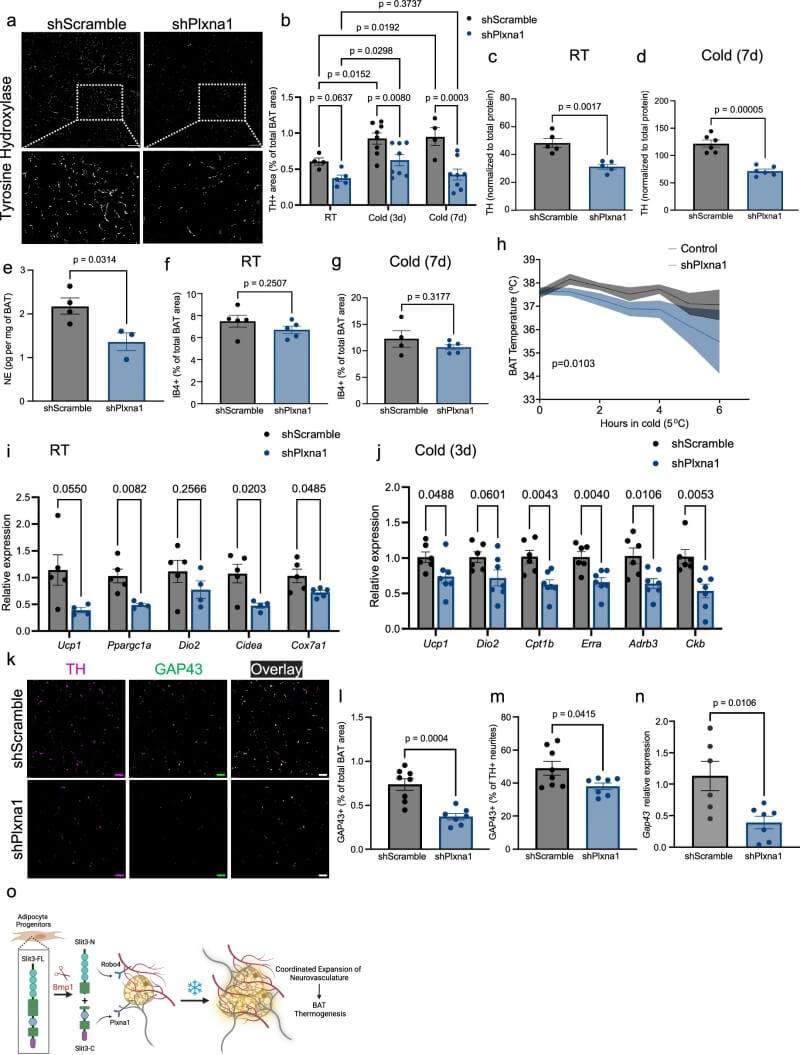 Fig.5 PLXNA1 is required for nerve remodeling and thermogenic responses in brown fat under cold conditions. (Serdan, et al., 2026)
Finally, the clinical relevance of this pathway was established using human fat biopsy cohorts from the Leipzig Obesity BioBank (LOBB). In insulin-sensitive individuals from the metabolically healthy versus unhealthy obese (MHUO) cohort, SLIT3 transcript levels in subcutaneous white adipose tissue (WAT) correlated positively with serum adiponectin and negatively with inflammatory macrophage content in omental visceral WAT. In a large cross-sectional cohort (CSC) of 1480 individuals, SLIT3 expression in omental visceral WAT correlated negatively with Hemoglobin A1c (HbA1c) levels and positively with circulating levels of the anti-inflammatory adipokine Omentin1, linking the SLIT3 pathway directly to improved metabolic health and insulin sensitivity in humans.
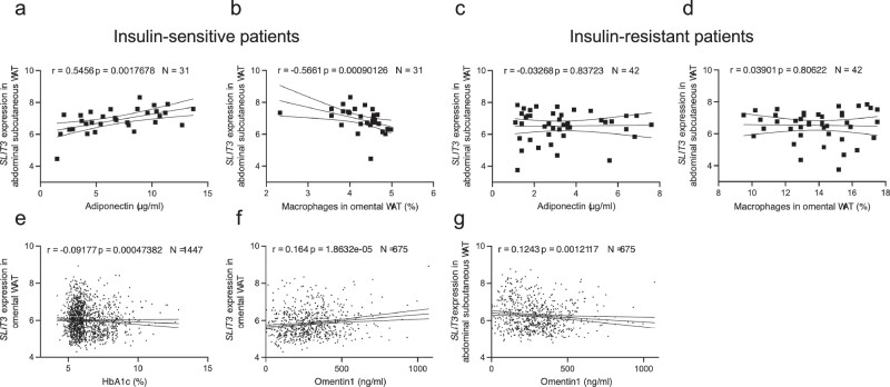 Fig.6 Higher SLIT3 expression is linked to healthier adipose tissue and lower inflammation in humans. (Serdan, et al., 2026)
These breakthrough findings hold profound implications for the global therapeutic landscape targeting metabolic disorders, obesity, and type 2 diabetes. By demonstrating that the neurovascular niche can be selectively expanded via distinct SLIT3 fragments, this study opens up an entirely new avenue for therapeutic engineering where researchers can bypass central nervous system pathways to directly drive peripheral energy expenditure. Therapeutics designed to mimic or stabilize SLIT3-C could potentially restore compromised sympathetic innervation in aging or diabetic adipose tissues, thereby reactivating dormant brown fat depots and enhancing global metabolic rates. Furthermore, discovering the BMP1-mediated cleavage mechanism provides a precise pharmacological target to fine-tune the balance between tissue vascularization and neural stimulation, shifting the focus of anti-obesity drug development from basic caloric restriction toward sophisticated tissue-remodeling strategies.
For research teams interested in exploring adipose neurovascular remodeling, metabolic disease pathways, or advanced tissue-clearing applications, Protheragen provides a comprehensive suite of preclinical CRO services, including single-cell transcriptomics, customized AAV vectors, high-resolution light-sheet imaging, and automated indirect calorimetry to accelerate your translational discoveries.
|
||
Reference
Copyright © Protheragen. All rights reserves.